Signalment:
2-month-old female
Belgian warmblood foal (
Equus
ferus caballusThe foal had a
sudden onset of diarrhea and fever (40°C). Blood ex-amination revealed elevated
liver values and abdominal ultrasound showed edema of the colon wall. Despite
treatment the foal died quickly.
Gross Description:
The foal was
admitted for necropsy and postmortem examination re-vealed a good nutritional
condition, and moderate dehydration. Petechial bleedings were noticed on the
pleura, the pericardium, the thymus, the splenic capsule and on the serosa of
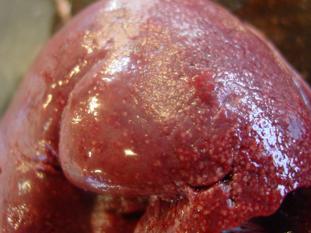
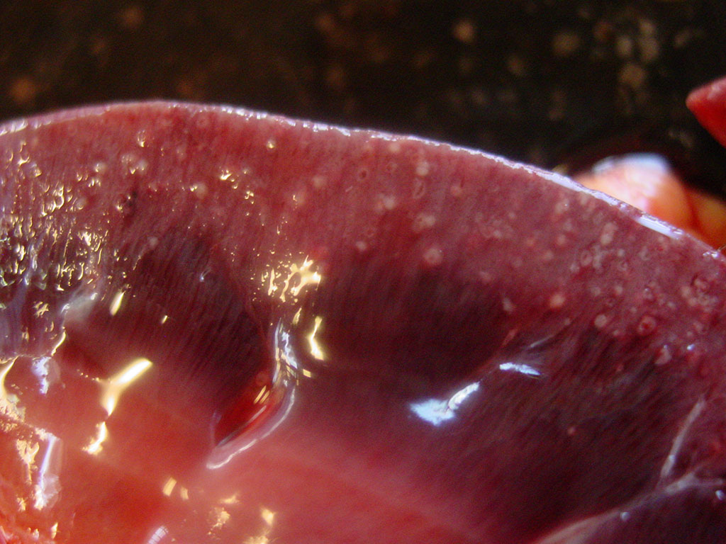
the intestine. The kidneys showed numerous cortical white to gray foci and
congestion of the medulla. The intestines were dilated with an edematous wall
and a mucoid gray content.
Histopathologic Description:
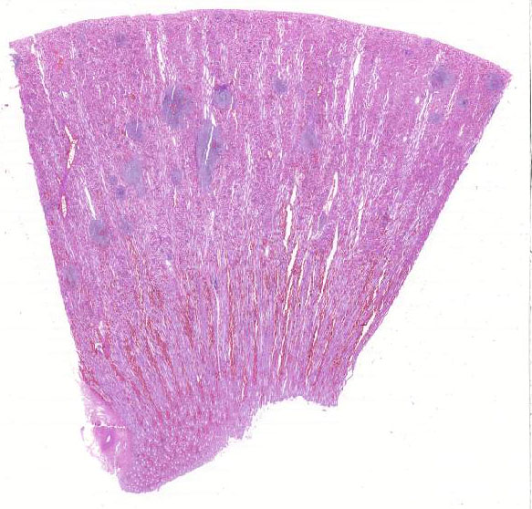
Kidney:
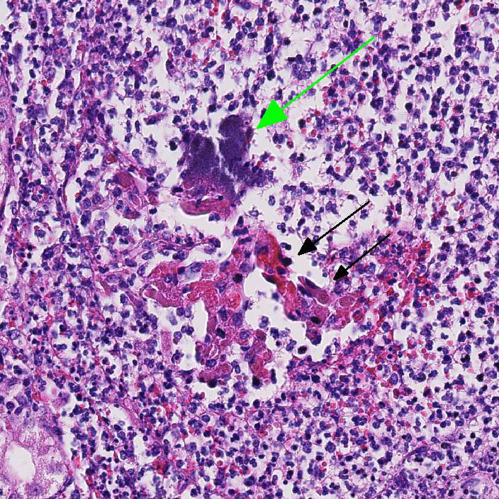
Randomly scattered within the cortex and occasionally extending into the medulla,
there are numerous embolic microabscesses (0.3-0.40 mm in diameter) that
regularly center on and efface glomeruli. These abscesses are composed of
abundant ne-crotic debris (karyorrhexis, karyolysis, and pyknotic nuclei),
admixed with many degenerate and non-degenerate neutrophils, fewer macrophages,
lymphocytes and plasma cells. Multifocally within these mic-roabscesses, there
are large colonies of basophilic coccobacilli (1x2 µm). Abscesses occasionally
extend into adjacent inter-stitium and tubules, with degeneration and necrosis
of tubular epithelium. There are multifocal areas of congestion, hemorrhage,
and fibrin thrombi within vessels.
Morphologic Diagnosis:
Kidney: Acute, severe, suppurative,
embolic nephritis with intralesional coccobacilli.
Lab Results:
Bacteriology
of the kidney: positive for
Actinobacillus equuli subsp. Haemolyticus
Parasitology of
the feces: positive for strongyles.
Condition:
Suppurative embolic neprhitis/Actinobacillus equuli
Contributor Comment:
Kidney: Acute, severe, suppurative,
embolic nephritis with intralesional coccobacilli. whether
such strains are common in-habitants of the equine gastrointestinal and respiratory
tracts.
5 Two subspecies of
Actinobacillus
equuli have been identified:
A. equuli subsp. equuli, and
A.
equuli subsp. haemolyticus.
1 The former appears to be
pathogenic, while the latters pathogenicity appears to be associated with its
expression of a repeats-in-structural-toxin (RTX) called Aqx, which is cytotoxic
for equine leukocytes.
1 Typically
actinobacillosis is a disease of newborn foals and the pathogenesis of the
infection remains speculative. Infection is probably acquired in utero, during
parturition, or shortly after birth as an um-bilical infection.
1
Death may occur due to fulminating septicemia. In foals that survive for
several days, microabscesses are seen in the kidney and other organs and a poly-arthritis
can be present. These micro-abscesses have an embolic origin and are
characterized by the presence of numerous, 1-3 mm, white pinpoint foci on the
cut surface throughout the renal cortex. Micro-scopically, glomerular
capillaries contain numerous bacterial colonies intermixed with necrotic debris
and extensive infiltrates of neutrophils that often obliterate the glomerulus.
4
The lesions in our
case are classic for
Actinobacillus equuli, and the foal was also
positive for strongyles. It has been post-ulated that migrating strongyle
larvae from the intestinal tract may play a role in infection.
JPC Diagnosis:
Kidney, cortex and
medulla: Nephritis, embolic, suppurative, acute, severe, with large colonies
of coccobacilli.
Conference Comment:
Despite some slide variability, the histopathologic appearance of this lesion
is a classic for suppurative and embolic nephritis caused by
Actinobacillus
equuli. This entity is the most common cause of suppurative and embolic
nephritis in young horses.
1 In pigs, embolic nephritis is most
commonly caused by
Erysipelothrix rhusiopathiae. In cattle,
Trueperella
pyo-genes from valvular endocarditis causes numerous septic emboli, which
shower the renal cortex causing randomly distributed microabscesses and
infarcts.
Corynebacterium pseudotuberculosis is most com-mon in sheep
and goats,
Pasteurella multocida in rabbits, and
Streptococcus
moniliformis in mice.
1,4 In dogs,
Prototheca zopfii
organisms have been identified as a common cause of embolic nephritis secondary
to systemic protothecosis.
1 Endotoxin expressed by gram-negative
bacteria and
Streptococcus sp. causes endothelial damage, vasculitis,
and bacterial emboli.
1,4 Most conference participants noted ectatic
tubules containing necrotic and sloughed tubular epithelial cells, fibrin,
hemorrhage, and proteinaceous fluid. Participants also noted occasional fi-brin
thrombi with colonies of coccobacilli within glomerular tufts, as well as
parietal cell hyperplasia secondary to the effects of endotoxin.
This case
illustrates the characteristic appearance of the large colony-forming
coccobacilli,
Actinobacillus equuli, in tissue section.
In addition to
discussing causes of embolic nephritis in other species, conf-erence
participants also reviewed other bacteria that form large colonies in tissue.
These bacteria are
difficult to distinguish from one another other on hematoxylin
and eosin stain (H&E), and require special stains or bacterial culture.1
Gram-positive large colony forming bacteria include: Staphylococcus, Streptococcus,
Actinomy-ces, and Corynebacterium spp.; while gram-negative large
colony forming bacteria include Yersinia and Actinobacillus spp.1,4
Several conference members men-tioned the acronym, YAACSS, as a helpful
mnemonic device to remember which bacteria form large colonies in tissue
section.
References:
1. Cianciolo RE, Mohr FC, Urinary system, In: Maxie MG, ed. Jubb,
Kennedy, and Palmers Pathology of Domestic Animals. Vol 2. 6th ed.
Philadelphia, PA: Elsevier Saunders; 2016: 432-433.
2. Frey J. The role of RTX toxins in host specificity of animal
pathogenic Pasteurellaceae. Vet Microbiol. 2011; 153:51-58. WSC
2012-2013.
3. Matthews S, Dart AJ, Dowling BA, et al. Peritonitis
associated with Actinobacillus equuli in horses: 51 cases.
Aus Vet J.
2001; 79:536-539.
4. Newman SJ: Urinary System. In: eds. McGavin MD, Zachary JF:
Pathologic Basis of Veterinary Disease. 5th ed. St. Louis, MO: Mosby
Elsevier; 2012:649.
5. Patterson-Kane JC, Donahue JM, Harrison LR. Septicemia and
peritonitis due to Actinobacillus equuli infection in an adult horse.
Vet
Pathol. 2001; 38:230-232.