Signalment:
11-year-old female spayed miniature schnauzer (Canis familiaris)
The dog presented for evaluation of a cataract and it was discovered that the dog is blind in this eye and has a probable retinal detachment. An enucleation was performed.
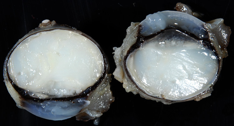
Gross Description:
The interior of the eye is filled with an opaque white material that obscures all internal structures.
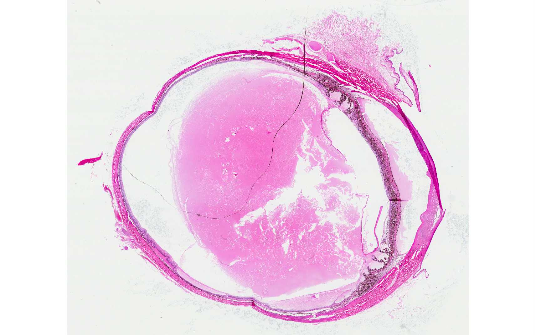
Histopathologic Description:
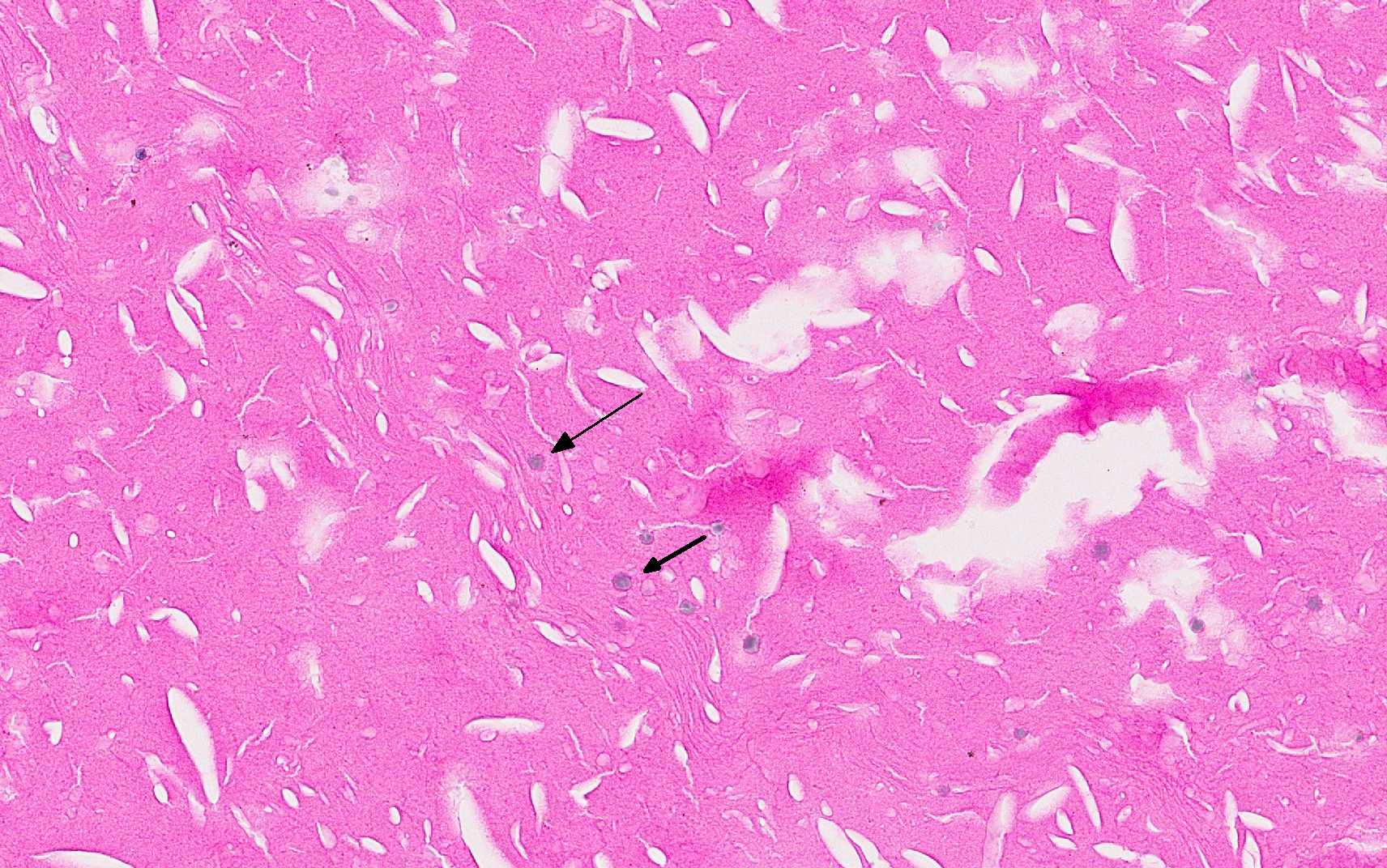
The vitreous and the anterior and posterior chambers are filled with a proteinaceous material containing many cholesterol crystals. A mild mixed inflammation of macrophages and lymphocytes is present at the margins of the fluid and a few macrophages are within the fluid. The retina has mild atrophy of the inner layers, a partial detachment, and a diffuse, mild infla-mmation of lymphocytes and plasma cells. The back of the iris is covered with a fibrous layer containing many lymphocytes. A few lens fibers are round, swollen and va-cuolated. The lens is not present in all slides.
Morphologic Diagnosis:
1. Cholesterolosis bulbi
2. Retinal atrophy, partial detachment and lymphoplasmacytic retinitis
3. Cataract
Lab Results:
Condition:
Contributor Comment:
Cholesterolosis bulbi is the accumulation of cholesterol within a degenerate vitreous humor that has become liquefied (syneresis). The condition may also involve the anterior and posterior chambers as in this case. It occurs in eyes that have suffered previous hemorrhage, in-flammation or other degenerative changes.2 The cause in this case is unknown but the cataract is a possibility.
JPC Diagnosis:
Conference Comment:
The cholesterol crystals are thought to be derived from the breakdown of intraocular erythrocytes and, may also be found within the anterior chamber.1,4 In people, cholesterolosis bulbi may be associated with a condition known as Coats+â-ó-é-¼-ä-ó disease, which includes retinal telangiectasis with intra and subretinal exudation.4 In cases of cholesterolosis in the absence of intraocular hemorrhage, it has been suggested that cholesterol crystals may enter eye through breaks in the retina and originate from the cholesterol rich subretinal fluid. Phacolysis has also been proposed as a source of cholesterol crystals.3
In addition to the proteinaceous, cholesterol-laden fluid filling most segments of the globe, conference participants noted retinal degeneration and atrophy, mild keratitis, uveal inflammation and choroidal edema. Fragmentation and fibrillation as well as cataractous changes were also described within the lens (not present in all sections), although the moderator cautioned against over-interpreting lens rupture due to the fragile nature of the lens and its tendency to fracture during sectioning. Specific retinal changes included retinal detachment, hypertrophy and hyperplasia of the retinal pigment epithelium, vacuolation of the nerve fiber layer, and degeneration of the ganglion cell and plexiform layers. The moderator commented on the rare nature of cholesterolosis bulbi, citing several potential underlying causes such as trauma and hemorrhage, although it should be noted that intraocular hemorrhage is a relatively common finding not specific to this uncommon condition. Asteroid hyalosis was also present and is typically characterized by the presence of 4-10um pale basophilic granules / globules within the posterior segment, consisting of lipids embedded in a matrix of calcium and phosphorus, and attached to the vitreous. The asteroid bodies appear as amorphous globules, are PAS positive and contain small birefringent crystals when viewed under polarized light. Diabetes mellitus is a risk factor in people for the development of asteroid hyalosis as are hypertension and atherosclerosis.
References:
1. Brodsky MC. Synchysis scintillans in a child. JAMA Ophthalmol. 2015; 133(7):e150793.
2. Grahn BH, Peiffer RL. Veterinary ophthalmic pathology. In: Gelatt KN, Gilger BC, Kern TJ, eds. Veterinary Ophthalmology. 5th ed. Hoboken NJ: Wiley-Blackwell; 2013:490.
3. Park, J, Lee H, Kim YK, Chae JD, Lee HJ. A case of cholesterosis bulbi with secondary glaucoma treated by vitrectomy and intravitreal bevacizumab. Korean J Ophthalmol. 2011;25(5):362-365.
4. Stacey AW, Borri M, Francesco SD, Antenore AS, et al. A case of anterior chamber cholesterolosis due to Coats+â-ó-é-¼-ä-ó disease and a review of reported cases. Open Ophthalmol J. 2016. Feb 29;10:27-32.
5. Yanoff M, Sassani JW. Ocular Path-ology. 6th ed. Mosby Elsevier; 2009:486-487.