Signalment:
10-year-old male Labrador retriever dog (
Canis familiaris).This dog had a clinical history of "CNS signs." The dog was a stray, and because it was acting "odd"
according to the property owner, the dog was submitted to the laboratory for rabies testing. The brain was removed
and the rest of the body was not examined.
Gross Description:
The cerebrum was distorted by an approximately 2 x 2.5 cm spherical, pale yellow-tan soft
friable mass. The mass was not encapsulated and was not connected to the meninges.
Histopathologic Description:
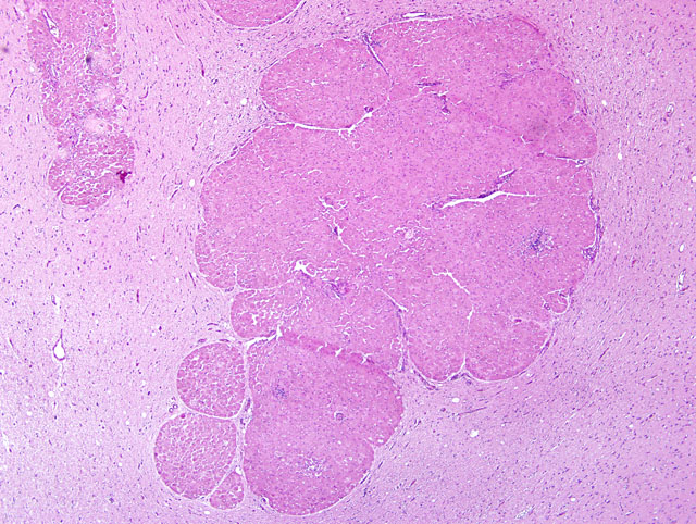
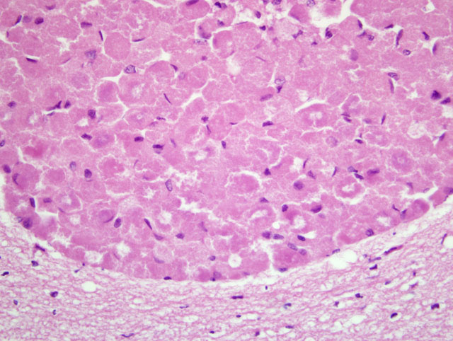
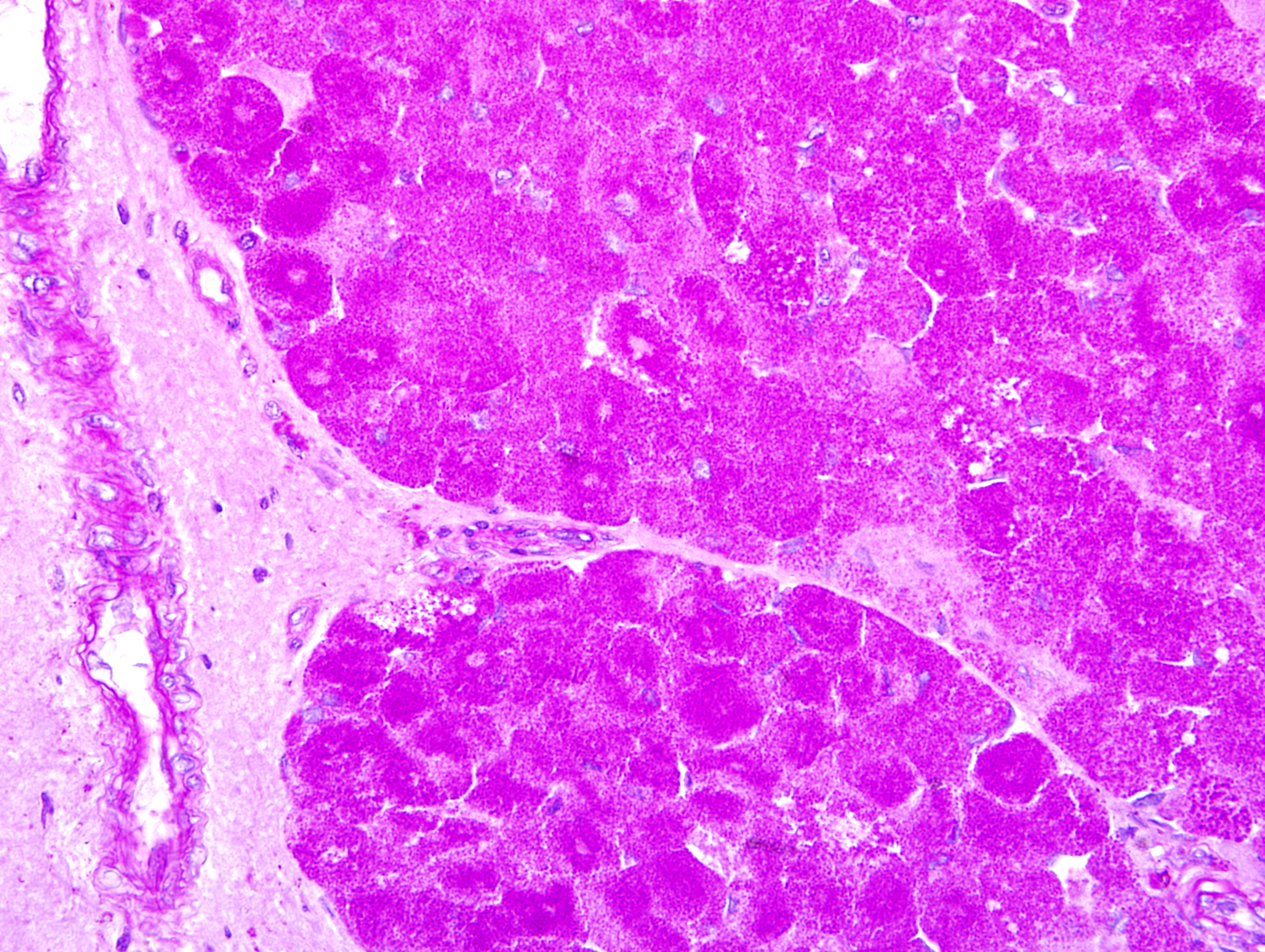
The meninges and adjacent neuropil have been infiltrated by numerous variablesized
nests of plump, round neoplastic cells. Individual nests are separated by a delicate fibrovascular stroma. The
neoplastic cells have finely granular eosinophilic cytoplasm, a central or eccentric, small, finely stippled nucleus
with one or two nucleoli. Mitotic figures are rare (not visible on all slides) and in some slides there are microfocal
lymphocytic perivascular cuffs within the tumor or the adjacent unaffected neuropil.
Morphologic Diagnosis:
Intracerebral granular cell tumor.
Lab Results:
Direct FA for rabies virus antigen was negative. An aerobic culture of the brain was positive
for
Pasteurella sp.
Condition:
Granular cell tumor
Contributor Comment:
In rats, granular cell tumors are the most common intracranial tumor.(5) In horses, this
type of tumor has only been reported in the lungs, where it may be an incidental finding.(4) In dogs, granular cell
tumors are most commonly located in the oral cavity.(2) Other, less frequently reported sites in dogs include the
heart, skin and brain.(1) Other species with reported granular cell tumors include cats(6) and ferrets.(5) Granular
cell tumors arising in the peripheral soft tissues of non-human animals are generally believed to originate from
Schwann cells.(1,4) The tumor cells usually contain diastase-resistant PAS-positive cytoplasmic granules.(1,3) A
definitive origin for granular cell tumors in the CNS of non-human animals has not been determined.
In humans, these tumors are histogenetically heterogeneous and occur both within the nervous system and in extraneural
locations.(3) The tumors in the CNS are believed to arise from astrocytes or pituicytes and tumors in extraneural
sites are believed to be of peripheral nerve origin (usually Schwann cells).(6)
In previously reported cases of intracranial granular cell tumors, the clinical signs have included blindness (when the
tumor entraps the optic nerves), seizures, ataxia, weakness, nystagmus, opistotonus, and proprioceptive deficits.
(1,3,5,6) Unfortunately, the history in this particular case was limited to "CNS signs" and "acting odd."
In this case, small perivascular cuffs of lymphocytes and less frequently neutrophils were present in the noninvolved
cerebrum and hippocampus (not included in most slides). In the cerebellum and hippocampus the
endothelial cells lining most blood vessels were plump and hypertrophic (reactive endothelial cells). Perivascular
cuffing associated with this type of tumor has been reported previously(1), and in this case we assume that the
lymphocytic perivascular cuffing is associated with the tumor while the neutrophils and reactive endothelium may
be in response to the presence of a
Pasteurella sp., believed to be an acute infection.
JPC Diagnosis:
Cerebrum: Granular cell tumor.
Conference Comment:
This case was reviewed in consultation with the AFIP Department of Neuropathology. It is
perplexing that while the gross description indicates that the neoplasm was not attached to the meninges,
microscopic evaluation reveals that the neoplasm, indeed, expands the meninges, often obliterating Virchow-Robin
space, and infiltrates the adjacent neuropil. As indicated by the contributors comments, the diverse list of examples
of granular cell tumors in various species suggests that the granular cell phenotype may develop in tumors that have
different cells of origin. While the histogenesis of granular cell tumors from various anatomic locations in several
animal species is known, the cell of origin of the cerebral granular cell tumor remains enigmatic.
Conference participants briefly discussed oncocytoma and laryngeal rhabdomyoma in the dog; these neoplasms
share histological similarities with granular cell tumors. Electron microscopy indicates that the prominent
cytoplasmic granules in granular cell tumors are lysosomes and autophagosomes.(1,3) In oncocytomas and
laryngeal rhabdomyoma, the cytoplasm is packed with mitochondria, accounting for the granules seen on H&E.(7)
References:
1. Barnhart KF, Edwards JF, Storts RW: Symptomatic granular cell tumor involving the pituitary gland in a dog: a
case report and review of the literature. Vet Pathol
38:332-336, 2001
2. Head KW, Else RW, Dubielzig RR: Tumors of the intestines.Â
In: Tumors in Domestic Animals, ed. Meuten DJ,
4th ed., p. 433. Iowa State Press, Ames, IA, 2002
3. Higgins RJ, LeCouteur RA, Vernau KM, Sturges BK, Obradovich JE, Bollen AW: Granular cell tumor of the
canine central nervous system: two cases. Vet Pathol
38:620-627, 2001
4. Kelley LC, Hill JE, Hafner S, Wortham KJ: Spontaneous equine pulmonary granular cell tumors: morphologic,
histochemical, and immunohistochemical characterization. Vet Pathol
32:101-106, 1995
5. Liu CH, Liu CI, Liang SL, Cheng CH, Huang SC, Lee CC, Hsu WC, Lin YC: Intracranial granular cell tumor in
a dog. J Vet Med Sci
66:77-79, 2004
6. Mandara MT, Ricci G, Sforna M: A cerebral granular cell tumor in a cat. Vet Pathol
43:797-800, 2001
7. Wilson DW, Dungworth DL: Tumors of the respiratory tract.Â
In: Tumors in Domestic Animals, ed. Meuten DJ,
4th ed., pp. 378-379. Iowa State Press, Ames, IA, 2002