Signalment:
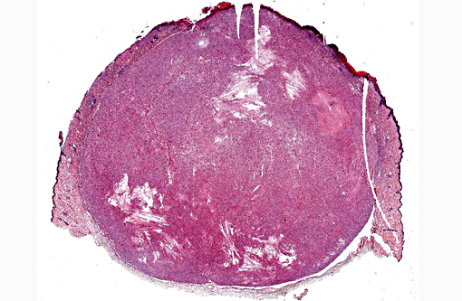
Gross Description:
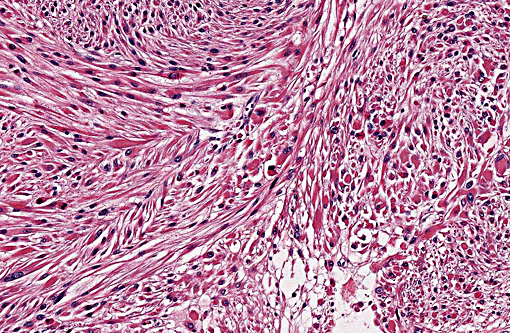
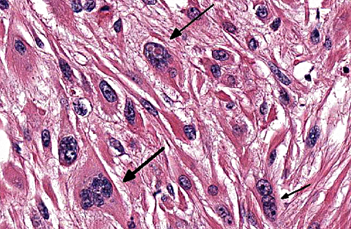
Histopathologic Description:
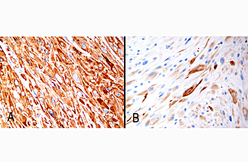
Immunohistochemistry (Figure 3-4A and 3-4B): Nearly all of the neoplastic cells have diffuse strong cytoplasmic immunoreactivity for smooth muscle actin and approximately 60% of the neoplastic cells have weak to moderate cytoplasmic immunoreactivity for desmin.
Morphologic Diagnosis:
Lab Results:
Condition:
Contributor Comment:
In ferrets, piloleiomyosarcomas most commonly arise from the head and trunk, with fewer cases from the limbs, and may have surface ulceration.(5,7) Histologically, despite being well demarcated, the neoplastic cells have significant nuclear pleomorphism, and reports vary regarding mitotic activity, which may be uncommon (1-2 per 10 high power fields)(7) or more frequent (≥ 2 per 10 high power fields).(5) The nuclear pleomorphism(7) or increased mitotic activity (mitotic index of ≥ 2)(5) are the foundation for the diagnosis of malignancy in these cases. Foci of lymphocytes may be seen at the periphery of the neoplasm(7) and areas of necrosis within the tumor may be identified,(5) as in the current case. Direct connection of the tumor with the adjacent arrector pili muscles may or may not be found,(5,7) and is not clearly present in this case. The majority of piloleiomyosarcomas in ferrets are positive for desmin and smooth muscle actin with immunohistochemistry(5,7) (Figure 3-4A and 3-4B). The prognosis for piloleiomyosarcomas in ferrets is good following complete surgical excision with no reports of metastasis and good long-term survival in those cases with follow up.(5,7) Follow up is not available for the present case.
In dogs and cats, general criteria to support a diagnosis of cutaneous leiomyosarcoma, as compared to a benign leiomyoma, include mitotic index of ≥ 1, evidence of invasion, and/or necrosis within the tumor.(1) In dogs and cats, piloleiomyomas and piloleiomyosarcomas are considered likely to be cured by complete excision, and local recurrence or metastasis of cutaneous leiomyosarcomas in general is uncommon.(1-3) In humans, leiomyosarcomas arising from the arrector pili muscles have a moderate local recurrence rate of 30% and metastasis is not reported.(7)
In conclusion, this case is a classic example of a piloleiomyosarcoma in a ferret and has many of the characteristic gross and histologic features reported with this entity.
JPC Diagnosis:
Conference Comment:
Cutaneous smooth muscle tumors can also arise from vascular smooth muscle and deep dermal smooth muscle of the genital area. Vascular origin smooth muscle tumors are termed angioleiomyoma/angioleiomyosarcoma. Regardless of origin, the tumors have similar histologic characteristics. Anatomic location can help differentiate these tumors, such as location in the genital area, or other features such as entrapment of hair follicles within the neoplasm, contiguity with erector pili muscle, or the presence/absence of a prominent vascular component may also be helpful.(5) Other neoplasms involving the skin of ferrets include mast cell tumors, basal cell tumors and neoplasms of apocrine sweat glands. Mast cell tumors are most common on the head, neck, shoulders and trunk but can occur at any location. Tumors of basal cell origin (most commonly sebaceous epithelioma of the skin can also occur at any location and are benign. The most common malignancy of the skin of the ferret are those of apocrine glands, which are most often seen around the prepuce and vulva, but may also be seen on the head and neck. Other less common tumors in the skin of ferrets include squamous cell carcinoma, cutaneous lymphoma, hemangioma/hemangiosarcoma, and fibroma/fibrosarcoma.(6)
References:
1. Cooper BJ, Valentine BA. Tumors of muscle. In: Tumors in Domestic Animals. Ames, IA: Iowa State Press; 2002:319-363.
2. Goldschmidt MH, Shofer FS. Uncommon skin tumors. In: Skin Tumors of the Dog and Cat. New York, NY: Butterworth Heinemann; 1998:291-295.
3. Liu SM, Mikaelian I. Cutaneous smooth muscle tumors in the dog and cat. Vet Pathol. 2003; 40:685-692.
4. Mialot M, Prata D, et al. Multiple progressive piloleiomyomas in a ferret (Mustela putorius furo): a case report. Vet Dermatol. 2010; 22:100-103.
5. Mikaelian I, Garner MM. Solitary dermal leiomyosarcomas in 12 ferrets. J Vet Diagn Invest. 2002;14:262-265.
6. Orcutt C, Tater K. Dermatologic diseases. In: Quesenberry KE, Carpenter JW, eds. Ferrets, rabbits, and rodents clinical medicine and surgery. 3rd ed. St. Louis, MO: Elsevier Saunders; 2012:127-130.
7. Rickman BH, Craig LE, Goldschmidt MH. Piloleiomyosarcoma in seven ferrets. Vet Pathol. 2001; 38:710-711.