CASE I: 1512649 (JPC 4083340)
Signalment:
3.5-month-old, male, goat (Capra aegagrus hircus).
History:
The goat had been examined by the submitting veterinarian, with an initial complaint of ataxia in its rear limbs by the owner. By the time of clinical evaluation, the kid was recumbent, blind and grinding its teeth. The owner of the flock reported that 4 additional young kids had a clinically similar disease during in spring 2015.
The kid was euthanized and presented for necropsy examination.
Gross Pathology:
No gross lesions were observed.
Laboratory Results:
The goat was deficient in copper (approximately one tenth the normal levels), with molybdenum concentrations over the normal range. Liver copper was measured at 2.6 ppm (Normal 25-150 ppm).
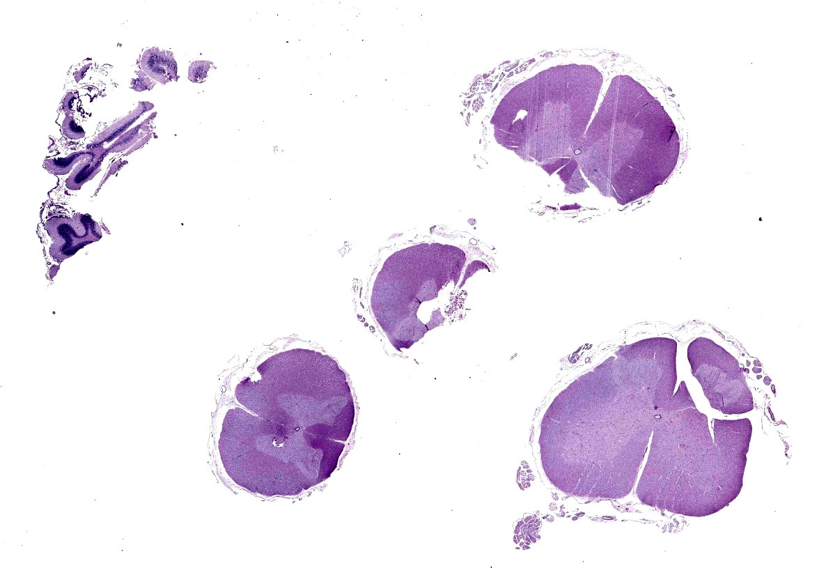
Microscopic Description:
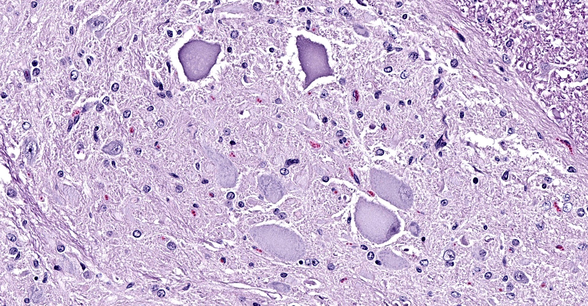
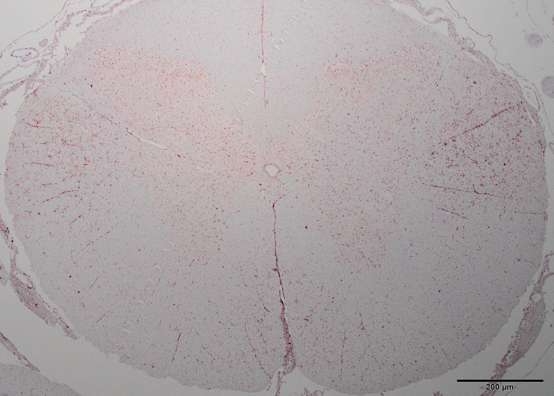
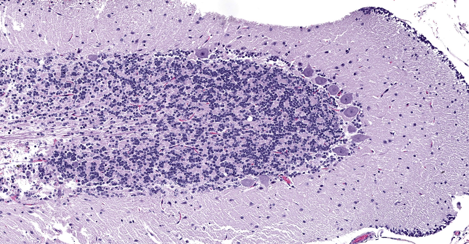
The spinal cord contained swollen, chromatolytic neurons that had lost Nissl substance, affecting Clarke's column and ventral horn neurons throughout the length of the cord. Pyknotic nuclei were rare. Similar changes were observed in cell bodies in the red and vestibular nuclei. These neurons retained cell-body phosphorylated neurofilaments. Spinal cord white matter degeneration was most severe in the lateral columns (spinocerebellar and spinomedullary tracts) and along the ventral median fissure, but was not easily visualized by myelin or neurofilament stains. The degree of tissue disturbance was more clearly demarcated with stains for microglia. CNPase did not demonstrate a reduction in oligodendrocyte numbers.
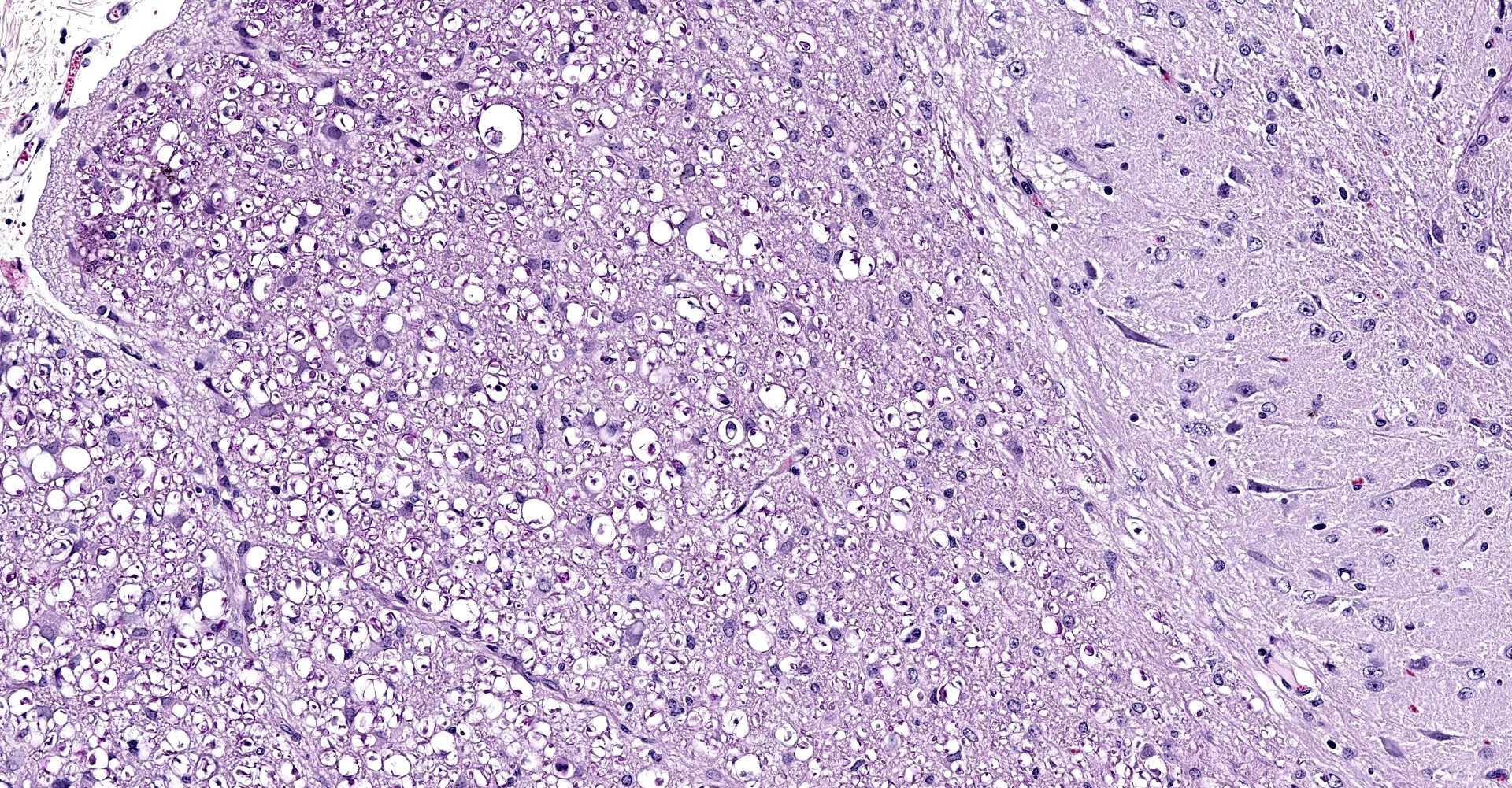
The kid had segmentally severe loss of cerebellar Purkinje and internal granular neurons in all folia, and occasional degenerating neurons were apparent with HE stain. Segmental cell loss was strikingly evident with calbindin immunohistochemistry. Some calbindin positive cells (Purkinje neurons) had dilated axons in the folial white matter near the cell body.
Contributor's Morphologic Diagnoses:
1. Purkinje cell
degeneration and loss (abiotrophy) with axonal swellings.
2. Neuronal degeneration, spinal cord.
Contributor's comment:
Copper is a metabolic cofactor of several enzymes, including mitochondrial cytochrome oxidase and superoxide dismutase in the central nervous system.4 Copper deficiency has a spectrum is microscopic lesions depending on timing and severity of disease. Ruminants are at particular risk, due to the potential of dietary copper binding to thiomolybdate complexes in the rumen. In one study, copper deficiency accounted for spinal ataxia in 13/23 goats.1 Most affected animals were less than 6 months of age, but other studies suggest goats up to 18 months of age can be affected.1,3
Lesions of copper deficiency are recognized world-wide in small ruminants, including goats.1,2,3,5 Number of cases varies widely between years, with variably severe clinical signs. In newborns, lesions consist of symmetrical necrosis of the cerebral white matter, sparing U fibers (swayback). Spinal cord changes of lateral white matter, termed demyelination in the literature, are described as concurrent axonal and myelin loss (axonopathy).
Cerebellar lesions are much more common in goats.3
"Swayback" has been recently reported in people, mostly women 50-70 years old who consumed excessive dietary zinc.4
Contributing Institution:
University of Missouri
Veterinary Medical Diagnostic Laboratory
vmdl.missouri.edu
JPC Diagnosis:
1. Spinal cord: Neuroaxonal degeneration, bilaterally symmetrical, moderate, with neuronal chromatolysis.
2. Cerebellum, Purkinje and granular cell neurons: Necrosis and loss, multifocal.
JPC Comment:
The contributor provides a concise review of copper deficiency, also known as hypocupremia, in ruminants.
Swayback and enzootic ataxia are two similar small ruminant central nervous system (CNS) diseases that occur as a result of a copper deficiency and are predominantly observed in small ruminants. Swayback is the term applied to the congenital form of disease which is primarily seen in neonatal lambs and rarely in kids. In contrast, enzootic ataxia has a delayed onset of clinical signs which typically manifest between one week and six months of age and affects both lambs and kids. Neonates with swayback are often stillborn, weak, unable to stand, or are ataxic while juveniles that develop enzootic ataxia are normal at birth but subsequently develop signs of disease characterized by incoordination, ataxia, and posterior paresis.2
Both forms of disease are due to insufficient dietary copper uptake, either by the dam while the fetus is in utero (swayback) or by the neonate following parturition (enzootic ataxia). Copper absorption from the diet is determined by the type of food, the mineral composition of the foodstuff, and the interactions between the food type and mineral composition. Copper is best absorbed from diets low in fiber, such as cereals, while fresh roughage is a poor source of copper. Due to the poor availability of copper in grass, primary copper deficiency most commonly affects grazing animals.3 In addition, elevations of dietary molybdenum, cadmium, sulfates, and other minerals or substances can interfere with copper absorption and utilization, resulting in secondary copper deficiency.1,2
As described by the contributor, copper is an essential cofactor utilized by multiple enzymes key to the function and development of the CNS, including the mitochondrial enzyme cytochrome oxidase. Dysfunction of this enzyme has been described in sheep and goats with swayback, resulting in decreased energy production and disruption of homeostasis within motor neurons.5
Both enzootic ataxia and swayback result in bilaterally symmetrical lesions of the grey and white matter within the cerebellum, brainstem, and spinal cord of both lambs and kids. Cerebral lesions are also observed in cases of swayback.5
Affected neurons are frequently swollen with chromatolysis, ranging from central chromatolysis with an eccentric nucleus to complete disappearance of Nissl substance with disruption and loss of nuclei (ghost cells). Other neurons are variably dense pink, and homogenous to fibrillar cytoplasm (due to accumulation of neurofilaments), have eccentric nuclei, and are variably necrotic.5 In cases of enzootic ataxia, affected neurons include the reticular formation in addition to the red and vestibular nuclei within the brainstem; the ventral, lateral, and less commonly the dorsal horns of the spinal cord; and Purkinjie cells within the cerebellum, which is also characterized by thinning of the granule cell layer and hypertrophic Bergmann glial cell processes within the molecular layer.5 In addition, Wallerian type degeneration is frequently observed in ventral spinal nerve rootlets and peripheral nerves.5 Respiratory stridor, likely due to degeneration of the recurrent laryngeal nerve may also be clinically observed.5
Affected regions of white matter are characterized by areas of pallor due to degeneration of myelinated axons. Specific regions of affected white matter affected by enzootic ataxia include the spinocerebellar tracts extending into the middle cerebellar peduncles and the the lateral and ventral funiculi of the spinal cord.5
While the above described lesions are characteristic, demonstration of low concentrations of copper in liver samples is diagnostic.1
References:
1. Allen AL, Goupil BA, Valentine BA. A retrospective study of spinal cord lesions of goats submitted to 3 veterinary diagnostic laboratories. Can Vet J. 2012;53:639-642.
2. Cordy DR, Knight, HD. California goat kids with a disease resembling enzootic ataxia or swayback. Vet Pathol. 1978;15:179-185.
3. Banton M, Lozano-Alcarcon F, Nicholson SS, et al. Enzootic ataxia in Louisiana goat kids. J Vet Diagn Invest. 1990;2:70-73.
4. Suttle,N.F. Copper imbalances in ruminants and humans: unexpected common ground. Adv Nutrition. 2012;6:666-674.
5. Wouda W, Borst,GHA, Gruys,E. Delayed swayback in goat kids, a study of 23 cases. Vet Quarterly.1986;8:45-56.