Signalment:  

10-month-old, intact male, pony, horse (< i>Equus ferus caballus).A ten month old male pony was submitted for necropsy with a history of poor growth and poor body condition. The submitting veterinarian suspected a congenital defect.


Gross Description:  

The animal was in poor body condition, with pale muscles and mucous membranes as well as marked subcutaneous edema. The liver was firm, tan to grey in color, and numerous cords of fibrous tissue coursed through the parenchyma. The entire left lobe and portions of the quadrate lobe were markedly atrophied (~50% of normal size). The right lobe was diffusely enlarged. Multifocally, fibrous villous proliferations were present on the serosal surface (perihepatitis villosa). Many adult trematodes, between 2-2.5 cm long, were evident within dilated and thickened bile ducts. The femoral metaphyseal bone marrow was reddened.


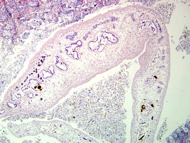
Histopathologic Description:

Liver: Liver sections reveal marked multifocal, mainly portal bridging fibrosis admixed with multifocal infiltrates of moderate numbers of eosinophils and large numbers of lymphocytes and plasma cells. There is marked bile duct hyperplasia. Large bile ducts contain an adult trematode characterized by a cuticle without spikes, a digestive tract, and vitellaria within a spongy parenchyma. Sections through adult trematodes show testicles with mature sperm, uteri with eggs, and also occasional miracidia larvae (morphologically consistent with Fasciola hepatica).


Morphologic Diagnosis:  

Marked diffuse chronic lymphoplasmacytic and eosinophilic hepatitis and (peri-)cholangitis with marked portal bridging fibrosis and intralesional adult trematode (morphologically consistent with Fasciola hepatica).


Lab Results:  

Clinical biochemistry: anemia, hypoalbuminemia.


Condition:  

Fasciola hepatica


Contributor Comment:  

In our experience (4 reports over a 21 year period), trematode infection is rare in equids in The Netherlands, and there are only a few reports of its prevalence in the literature. The reported prevalence of infection of horses in Europe ranges from approximately 1% in central Europe to 77% of horses from Ireland and the United Kingdom.(4) A German study found that between 38 and 71% of horses and 11% of donkeys were positive for the presence of eggs in their feces, while a similar Turkish study reported that the feces of 4.7% of equids were positive for eggs.(2,5)

Fascioliasis is common in cattle and sheep. The life cycle of Fasciola hepatica includes a Lymnead snail as an intermediate host. The snail is found in damp environments, such as marshy ground, ponds, and along the banks of slow moving streams. The miracidia of Fasciola hepatica, which are ingested along with the feces of the final host, infect the snail by burrowing through its body wall. Once within the snail, the larvae pass through a number of developmental stages including a sporocyst, three generations of rediae, and finally a cecarial stage which leaves the snail and encysts on vegetation to become a metacercaria. The infective metacercariae are ingested by the final host and excyst within the duodenum and migrate to the liver. Adult flukes reside in the bile ducts.(3)

In comparison to cattle and sheep, equids appear to mount an, as yet poorly understood, early immunological challenge to infection, which leads to the destruction, immobilization or developmental retardation of the larval flukes, the result being that only a few trematodes reach maturity in the bile ducts of equids.(2)


JPC Diagnosis:  

Liver: Cholangiohepatitis, proliferative and fibrosing, chronic and eosinophilic, diffuse, marked with hepatocellular loss, dark brown anisotropic pigment, and few trematode eggs and adults.


Conference Comment:  

Based on the histologic findings, a reasonable differential diagnosis list would include Fasciola hepatica, Fasciola gigantica, and Dicrocoelium dendriticum. All three of these trematodes reside in the bile ducts of affected animals; Fascioloides magna inhabits the liver parenchyma and therefore would be an unlikely etiologic agent. Speciation of trematodes in histologic samples is nearly impossible. With the exception of F. magna, the histologic lesions are similar, and thus identification of flukes at gross necropsy is more reliable, as the morphology can be more adequately characterized. To that end, F. hepatica is approximately 2.5 cm long and leaf shaped; F. gigantic is double or triple the size of F. hepatica; D. dendriticum is 0.5-1cm long and lancet-shaped; and F. magna is 8 cm in length.(3)

Given the limited clinical information and history provided to participants, i.e. species of animal affected, most favored D. dendriticum as the causative agent. In addition to infecting horses, this trematode also infects ruminants, pigs, dogs and cats. Fasciola hepatica and F. gigantica primarily affect sheep and cattle. Wild cervids are the natural host for F. magna, though it can infect cattle and sheep. Other trematodes infecting domestic animals include: Eurytrema pancreaticum and E. coelomaticum in ruminants; Opisthorchis viverrini in cats and dogs; and Pseudamphistimum truncatum, Metorchis spp., Parametorchis complexus, Concinnum procyonis, and Platynosum fastosum in cats and dogs.(1)

Two possible sequelae to hepatic trematodiasis are black disease in sheep and bacillary hemoglobinuria in sheep and cattle. Black disease occurs when anaerobic, necrotic foci in the liver resulting from F. hepatica migration allow ingested Clostridium novyi spores in the liver to germinate. Once active, C. novyi elaborates a necrotizing beta toxin and hemolytic phospholipase C, and together, these toxins produce large areas of coagulative necrosis in the liver resulting in severe edema, congestion, hemorrhage and death, often with no premonitory signs. The pathogenesis of bacillary hemoglobinuria, caused by Clostridium haemolyticum, is similar to black disease with respect to the fluke involved, germination of spores, and toxin production. The toxins of C. haemolyticum produce intravascular hemolysis with associated anemia and hemoglobinuria.(1)


References:

1. Cullen JM. Liver, biliary system and exocrine pancreas. In: McGavin MD, Zachary JF, eds. Pathologic Basis of Veterinary Disease. 4th ed. St. Louis, MO: Elsevier; 2007:435-436.
2. Eckert J. Helminthosen der Equiden. In: Rommel M, Eckert J, Kutzer E, K+�-�rting W, Schnieder T, eds. Veterin+�-�rmedizinische Parasitologie. 5th ed. Parey; 2000:353-354.
3. Stalker MJ, Hayes MA. Infectious diseases of the liver. In: Maxie MG, ed. Jubb, Kennedy and Palmers Pathology of Domestic Animals. 5th ed., vol. 3. Philadelphia, PA: Elsevier Ltd; 2007:354-356, 359-362.
4. Wintzer HJ, Kraft W. Parasit+�-�re Krankheiten des Pferdes. In: Wintzer HJ, ed. Krankheiten des Pferdes. 3th ed. Parey; 1999:229.
5. Uslu U, Guclu F. Prevalence of endoparasites in horses and donkeys in turkey. Bull Vet Inst Pulawy. 2007;51: 237-240.


Click the slide to view.


1-1. Liver


Back | VP Home | Contact Us |