WSC 22-23:
Conference 8:
CASE III:
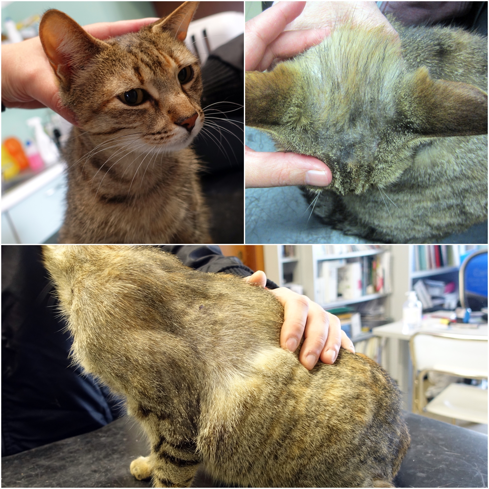
Signalment:
5-year-old spayed female European Shorthair cat (Felis catus)
History:
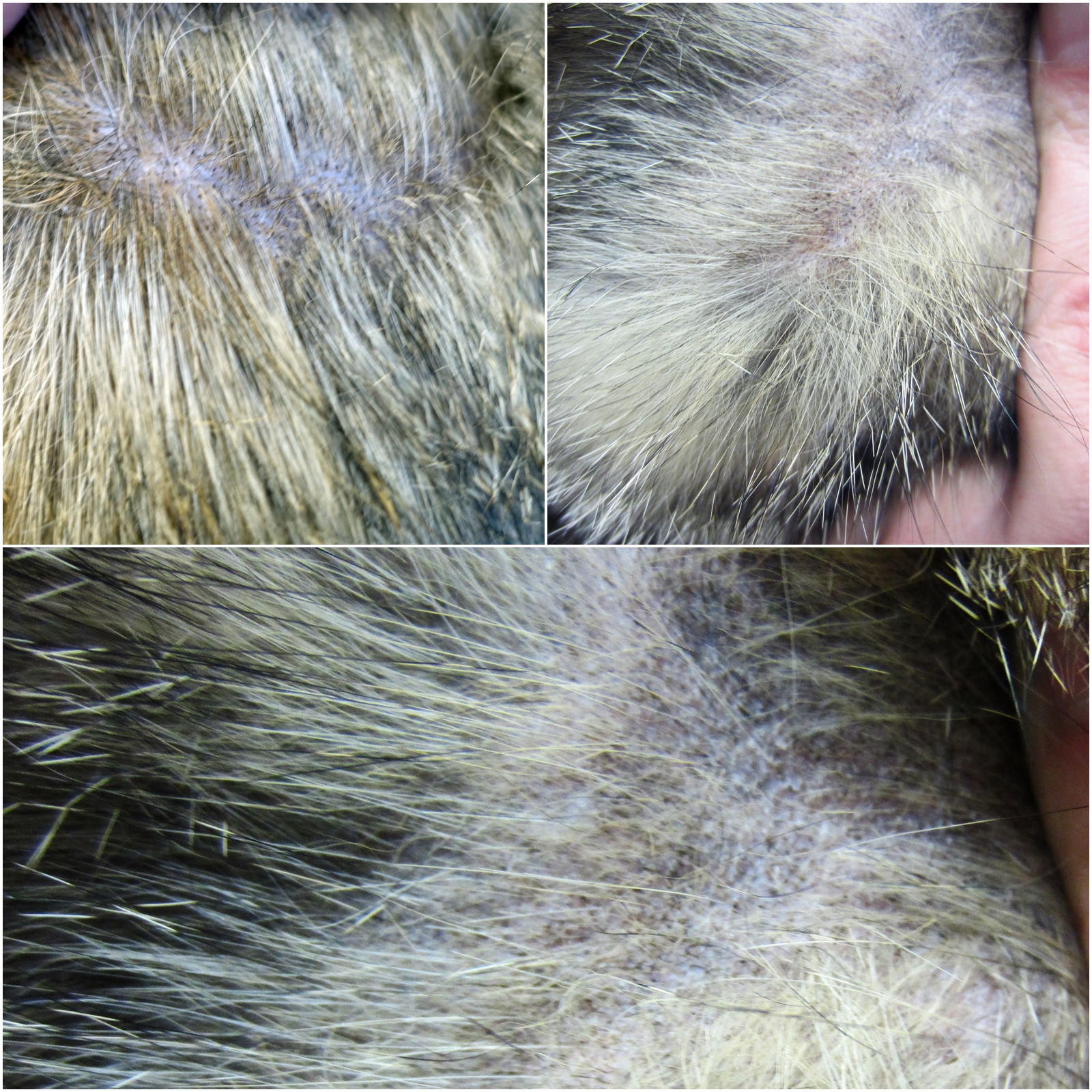
The cat had a chronic history of diffuse exfoliative dermatitis with multifocal alopecia, in particular in the region of head, trunk and limbs with scaling, hair casts, flakes, easy detachable hairs and moderate itching.
Based on clinical history the main differential diagnoses were: demodicosis, dermatophytosis, exfoliative dermatitis (thymoma associated or not), and sebaceous adenitis.
Gross Pathology:
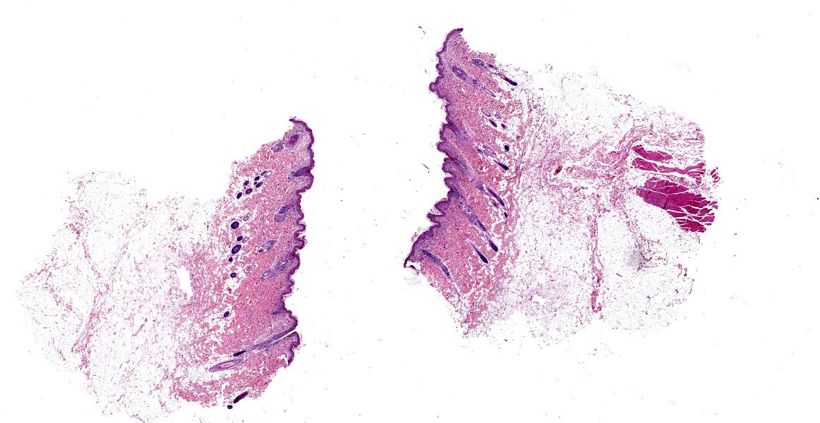
Four skin biopsies (8 mm punch and surgical scissors excision) were performed at the level of the neck (right side), costate, right thigh.
Laboratory Results:
Microbiology: negative.
Deep skin scraping: negative.
PAS stain: negative.
Microscopic Description:
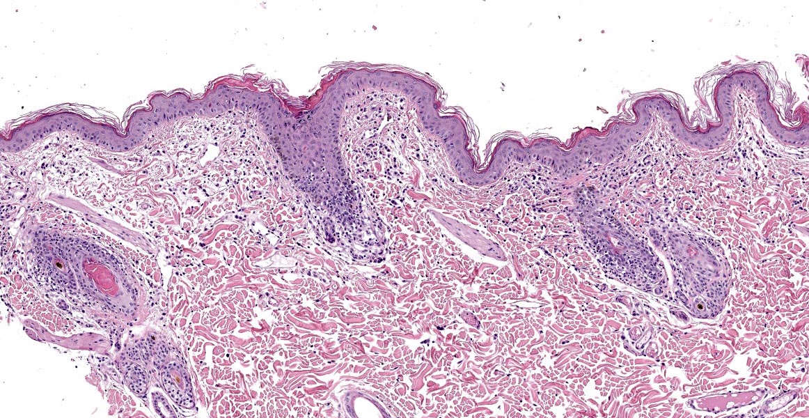
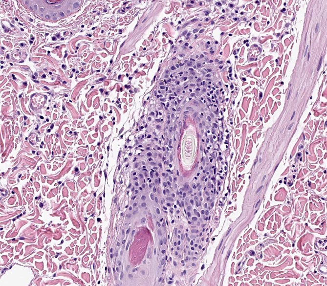
Haired skin. Haired skin is affected by a moderate inflammatory change involving hair follicles and obscuring sebaceous glands. Follicular walls are transmurally infiltrated, mainly in the region of isthmus and extending to the other portions, by a high number of lymphocytes and a lower number of macrophages and rare neutrophils. Keratinocytes of the outer root sheath frequently have vacuolated cytoplasm and rarely are shrunken, hypereosinophilic with pyknotic nuclei (apoptosis). In the superficial dermis a mild perivascular to interstitial inflammatory infiltrate composed, in a similar amount, of mast cells, lymphocytes, plasma cells and eosinophils is present associated with scant interstitial edema.
Contributor’s Morphologic Diagnoses:
Haired skin:
-moderate chronic lymphocytic mural folliculitis with loss of sebaceous glands
-mild perivascular to interstitial superficial dermatitis lymphoplasmacytic, with mast cells and eosinophils
Contributor’s Comment:
Histopathological findings are consistent with a sebaceous adenitis associated with mural folliculitis. Sebaceous adenitis is an uncommon disease in dogs and rabbits and extremely rare in cats.
Sebaceous adenitis in dogs, although it has idiopathic origin, in standard poodles and akitas can be inherited through an autosomal recessive gene with variable expression. Hypothesis made for its pathogenesis include a cornification disorder, a cell-mediated destruction of sebaceous glands, an anatomic defect in sebaceous glands leading to lipid leakage and a foreign body response, and a metabolic defect in lipid metabolism leading to a cornification abnormality and sebaceous gland destruction.1,3,6
Histology of canine sebaceous adenitis varies depending on the duration of disease and the breed affected. In the early stages histological lesions are characterized by a granulomatous to pyogranulomatous inflammation targeting the sebaceous glands while, in the chronic stage, which are more frequently submitted to laboratories, appears in an almost total loss of sebaceous glands. Severe epidermal orthokeratotic hyperkeratosis and follicular keratosis are invariably present. In Standard Poodles the late stage of disease may be characterized by follicular atrophy and an absence of sebaceous glands, while in Vizslas inflammation is more consistently prominent at all stages of the disease and the main differential diagnosis, in this case, is leishmaniasis.1,3
In rabbits, in addition to what is described in dogs, an interface dermatitis and an interface folliculitis are often present, suggesting that sebaceous adenitis may be only one aspect of a more generalized disorder.10
In cats, it is rarely described and it occurs as a chronic progressive disease with non-itchy scaling, crusting, alopecia and skin depigmentation in regions of the face, cervical and trunk which are the same features found in the case described except for pruritus. The latter has been described to be related to secondary infections and /or Malassezia spp. Overgrowth.2,4,6
In our case the definitive diagnosis was established histologically. The main lesions were the loss of sebaceous glands and a mural folliculitis, prominent in the isthmic region. The former is frequently described in sebaceous adenitis in different species; the latter is a reaction pattern of cats that has been associated with several diseases, included sebaceous adenitis but also with allergic dermatoses.7 In this case, once dermatophytosis and demodicosis were excluded with special stains, negative cultures and deep skin scraping, histological lesions associated with clinical presentation led to the diagnosis of sebaceous adenitis with mural folliculitis. Treatments proposed include cyclosporin therapy; in one case, topical fatty acid supplementation was also reported.2,4 Our case improved quickly with a cyclosporin therapy.
Contributing Institution:
San Marco Veterinary Clinic and Laboratory
Pathology division
Viale dell’Industria 3, 35030
Veggiano (PD), Italy
https://www.clinicaveterinariasanmarco.it/
JPC Diagnosis:
- Haired skin: Sebaceous adenitis, lymphocytic, diffuse, marked with sebaceous gland loss and multifocal mural folliculitis.
- Haired skin: Dermatitis, mastocytic and eosinophilic, superficial and perivascular, mild.
JPC Comment:
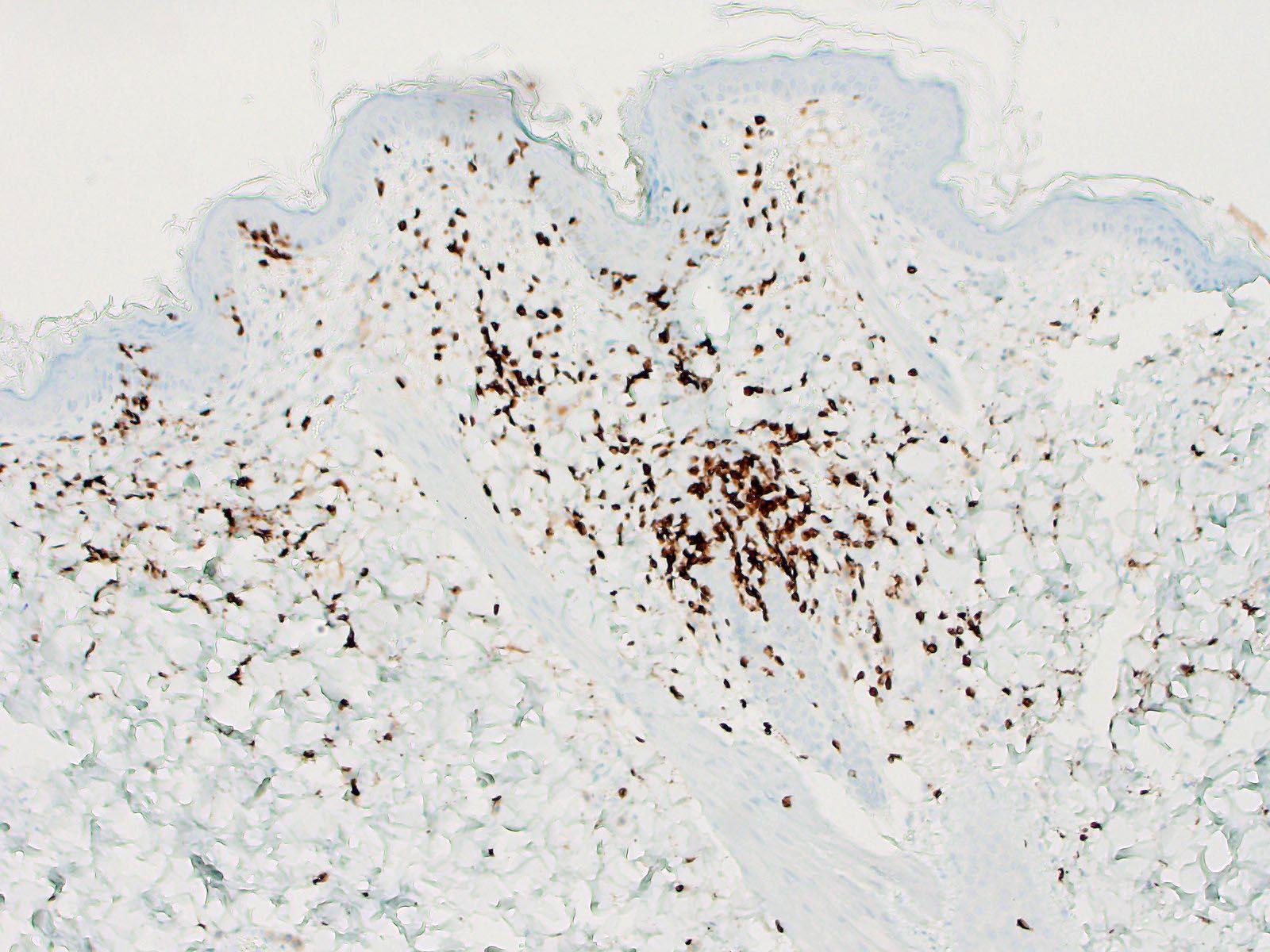
One hallmark gross lesion of sebaceous adenitis is the follicular cast, as demonstrated in figure 3-2, which appear grossly as flaky concretions surrounding the bases of hair shafts, though this is less common in short haired dogs.9
As the contributor mentioned, a main gross differential in this cat is exfoliative dermatitis. Histologically, the mural folliculitis of exfoliative dermatitis is focused on the infundibulum and isthmus and is frequently accompanied by secondary sebaceous adenitis or absence of sebaceous glands, which may mimic sebaceous adenitis.9 Exfoliative dermatitis can be differentiated, however, by its variable lymphocytic interface dermatitis with hydropic degeneration of basal keratinocytes, suprabasilar keratinocyte apoptosis, and orthokeratotic hyperkeratosis.9
Standard poodles are predisposed to a sebaceous adenitis, in addition to a few other autoimmune diseases, including primary hypoadrenocorticism. This high incidence of these two diseases have been linked to an artificial genetic bottleneck which occurred in the mid 20th century resulting from the closed breed registry and intensive breeding of a handful of show-winning dogs, including Sir Gay, Wycliffe Jacqueline, Bel Tor, Carillon, and a few other dogs with equally grandiose names. 5 A study of pedigrees and DLA (MHC) markers in standard poodles from the US, Canada, and Europe showed that this mid-century bottleneck decreased genetic diversity within 70% of the standard poodle population and concentrated the genetic polymorphisms underlying sebaceous adenitis and primary adrenocorticism. 5 Geneticists recommend careful mate selection based on pedigree and genetic testing to improve the diversity within the breed and decrease incidence of disease within this breed.5 As there is no currently available DNA test for sebaceous adenitis, the Orthopedic Foundation for Animals (OFA) recommends histologic evaluation of biopsies every 1 to 2 years in breeding animals greater than 12 months of age to assist in removing affected animals from the breeding population.8
References:
- Bardagì M, Fondevila D, Zanna G et al. Histopathological differences between canine idiopathic sebaceous adenitis and canine leishmaniosis with sebaceous adenitis. Veterinary Dermatology. 2010; 21: 159-165.
- Glos K, von Bomhard W, Bettenay S et al. Sebaceous adenitis and mural folliculitis in a cat responsive to topical fatty acid supplementation. Veterinary Dermatology. 2016; 11:57-60.
- Gross TL et al. Skin diseases of the dog and cat. Second edition. Blackwell Publishing; 2005.
- Noli C, Toma S. Three cases of immune-mediated adnexal skin disease treated with cyclosporine. Veterinary Dermatology. 2006;17: 85-92.
- Pederson NC, Brucker L, Tessier NG, et al. The effect of genetic bottlenecks and inbreeding on the incidence of two major autoimmune diseases in standard poodles, sebaceous adenitis and Addison’s disease. Canine Genetics and Epidemiology. 2015 (2):14-32.
- Possebom J, de Farias MR, de Assunção DL et al. Sebaceous Adenitis in a Cat. Acta Scientiae Veterinariae. 2015; 43:1-5.
- Rosenberg AS, Scott DW, Hollis N et al. Infiltrative lymphocytic mural folliculitis: a histopathological reaction pattern in skin-biopsy specimens from cats with allergic skin disease. Journal of Feline Medicine and Surgery. 2010; 12: 80-85.
- Sebaceous adenitis. OFA. Accessed October 12, 2022. https://ofa.org/diseases/other-phenotypic-evaluations/sebaceous-adenitis/
- Welle MW, Linder KE. The Integument. In: Zachary JF, ed. Pathologic Basis of Veterinary Disease. Louis, MO: Elsevier. 2022; 1246-1247,1261.
- White SD, Linder KE, Shultheiss P et al. Sebaceous adenitis in four domestic rabbits. Veterinary Dermatology. 2000; 11: 53-60.