Signalment:  

Twelve-week-old, female, rabbit, (Oryctolagus cuniculus).Per contributor: “Rabbits have been dying around 12 weeks of age. No previous clinical signs. Another rabbit had liver abscesses as well. Rule out Pasteurella, Staph, tularemia? Yellow and white abscesses.”


Gross Description:  

Tissue (liver) is submitted fixed in neutral buffered formalin. The liver parenchyma contains numerous, variably-sized, 0.1-0.2cm in diameter, firm, tan, elliptical nodules.


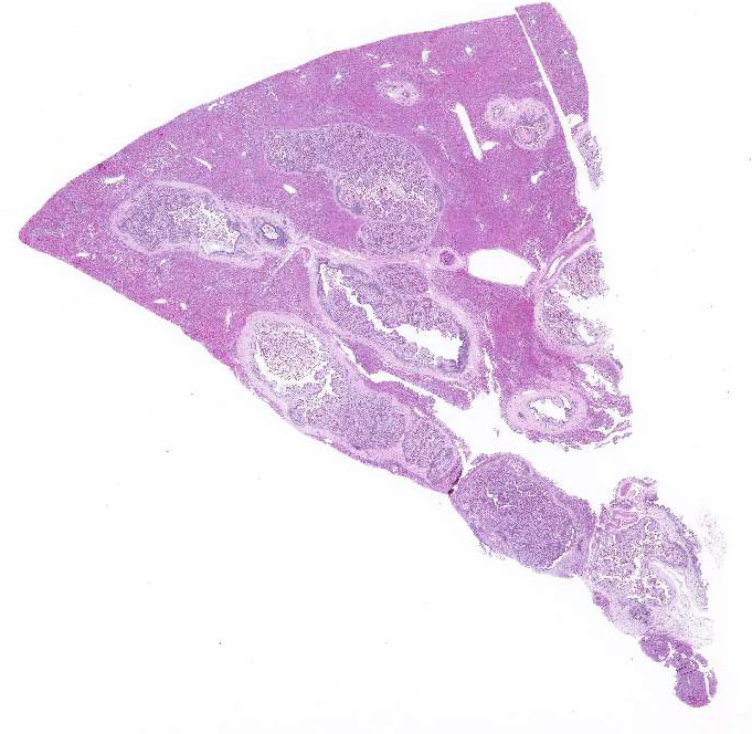
Histopathologic Description:

Significant microscopic lesions are focused on portal regions. Bile ducts are markedly dilated, tortuous and encased in abundant fibrous connective tissue. The biliary epithelium is markedly hyperplastic, usually lining papillary-type projections that fill the lumen of the dilated ducts. The hyperplastic biliary epithelium contains conspicuous sexual stages of protozoal micro- and macrogametes with less conspicuous asexual developmental stages. The bile duct lumens contain myriad oocysts. The fibrous connective tissue supporting the papillary projections and surrounding the dilated bile ducts contains infiltrates by primarily lymphocytes and plasma cells.  Elsewhere in the sections, the hepatocytes exhibit mild, zonal (centrilobular) atrophy.


Morphologic Diagnosis:  

Liver: Marked to severe, chronic, proliferative cholangitis, lymphoplasmacytic with portal and bridging portal fibrosis and myriad, intralesional protozoa consistent with Eimeria stiedae.


Lab Results:  

N/A


Condition:  

Liver cocciciosis/Eimeria stiedae


Contributor Comment:  

Hepatic coccidiosis (Eimeria steidae) can occur in either wild or domestic rabbits and represents an important cause of mortality in commercial rabbitries.4 Although infections can be subclinical or even incidental findings at necropsy, significant infections result in clinical signs of anorexia, lethargy, diarrhea, distended abdomen and poor weight gain. In this case, the young rabbits were dying around 12-weeks of age without premonitory clinical signs. Because of the acute mortality and gross lesions, the attending veterinarian was concerned about

Nonpathogenic

Slightly Pathogenic

Pathogenic

Highly Pathogenic

E. coecicola (appendix, sacculus rotundus, Peyers patches)

E. perforans (entire small intestine)

E. media (duodenum to jejunum)

E. intestinalis (lower jejunum and ileum)


E. exingua (emall intestine

E. magna (jejunum and ileum)

E. flavescens (small intestine and cecum)


E. vejdovskyi (ileum)

E. piriformis (colon)


E. irresidua (jejunum and ileum


differential diagnosis of tularemia in this case. There is some overlap of hepatic gross lesions and tularemia is not uncommon in this geographic region (Oklahoma). Concurrent infection by tularemia and hepatic coccidiosis has been previously described2; therefore, these gross liver lesions should always be microscopically investigated for possible tularemia, especially in wild rabbits.

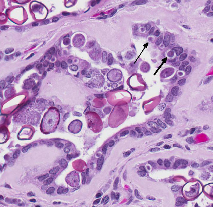
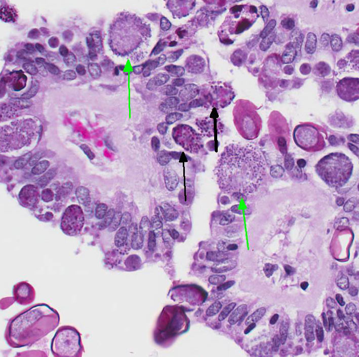
After ingestion of sporulated oocysts, sporozoites penetrate the duodenal mucosa and eventually spread to the liver by lymphatic and hematogenous routes.4 Upon reaching the liver, the sporozoites invade the biliary epithelium and enter the typical coccidian life cycle of schizogony, then gametogony with production of oocysts that are released into the bile ducts and passed into the intestine.4 The prepatent period is approximately 15-18 days.4

The severity of disease appears to be related to the initial dose level of the oocyst inoculum.3 Antemortem diagnosis is typically made by clinical signs and demonstration of oocysts in the feces. Post-mortem findings of the proliferative biliary lesions and histological identification of the organisms are pathognomonic for the disease.4 Control of hepatic coccidiosis can be achieved with improvement of hygienic conditions, particularly removal of fecal material containing oocysts before they finish sporulation.3 As it is probably impossible to remove all oocysts, control is augmented by prophylaxis with different anticoccidian drugs.1,3 It should be noted that some are better than others and drugs that are effective in controlling chicken coccidiosis are not always efficacious in rabbits.


JPC Diagnosis:  

Liver: Cholangitis, proliferative, lymphoplasmacytic, chronic, diffuse, marked, with portal and bridging fibrosis and numerous intraepithelial coccidial schizonts, gamonts, and oocysts rabbit, Oryctolagus cuniculus.


Conference Comment:  

The contributor provides a concise review of epidemiology, life cycle, and lesions associated with hepatic coccidiosis in rabbits. Conference participants readily identified the numerous schizonts containing merozoites, micro- and macrogametocytes, and micro- and macrogametes the biliary epithelium and oocytes in the lumen of the markedly proliferative bile ducts. Eimeria sp. and Isospora sp. are relatively host specific coccidian parasites of the phylum Apicomplexa that typically affect the mucosal and ductular epithelial cells of the gastrointestinal tract mucosa in many different animal species.3,4 Readers are encouraged to review for a list of important coccidian parasites of veterinary importance.

Eimeria sp. typically cause subclinical disease but can cause serious illness in young and immunosuppressed lagomorphs. Over 11 species of coccidian parasites have been described in rabbits and E. stiedae is the only coccidian parasite found in the liver and biliary epithelium .3,4 As mentioned by the contributor, the rabbit is infected by ingestion of sporulated oocysts in the feces with sporozoites invading the duodenal mucosa. Sporozoites have been documented in the regional lymph nodes within 12 hours, in bone marrow within 24 hours, and in the liver within 48 hours via hematogenous spread by infecting mononuclear cells.4 Rabbits with hepatic coccidiosis are often co-infected with intestinal Eimeria sp.  Intestinal Eimeria sp. target specific segments of the intestinal tract in rabbits and are divided into four groups based on pathogenicity in the following table:

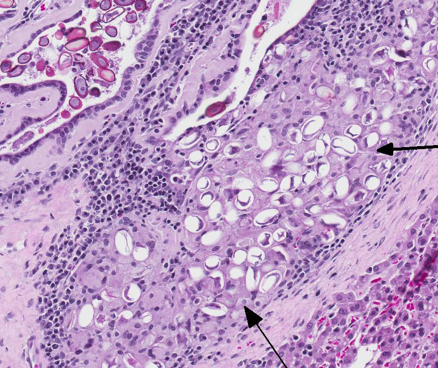
Table adapted from Pakandl M3 and Percy DH and Barthold SW4 The marked proliferation of the biliary epithelium, seen in this case, is secondary to destruction and regeneration of the bile duct epithelium with extensive hyperplasia of the ductular epithelium. In addition, biliary outflow may be obstructed by numerous oocysts within the lumen of the bile duct resulting in cholestasis and further distention of the bile duct.4

Conference participants discussed whether this case represents cholangiohepatitis with inflammation extending into the hepatic parenchyma or simply cholangitis with secondary mild centrilobular atrophy of hepatocytes and replacement by bridging fibrous connective tissue. Conference participants overwhelmingly agreed with the contributor that while the markedly dilated bile ducts compress the adjacent hepatic parenchyma, there is no disruption of the hepatic limiting plate; thus favoring the morphologic diagnosis of lympho-plasmacytic cholangitis.


References:

1. Cam Y, Atasever A, Eraslan G, Kibar M Atalay O, Beyaz L, Inci A, Liman BC. Eimeria stiedae: experimental infection in rabbits and the effect of treatment with toltrazuril and ivermectin. Exp Parasitol. 2008; 119:164-172.
2. Kim DY, Reilly TJ, Schommer SK, Spagnoli ST. Rabbit tularemia and hepatic coccidiosis in a wild rabbit. Emerg Inf Dis. 2010; 16:2016-2017.
3. Pakandl M. Coccidia of rabbit: a review. Folia Parasitol. 2009; 56:153-166.
4. Percy DH, Barthold SW. Rabbit. In: Pathology of Laboratory Rodents and Rabbits. 4th ed. Ames, IA: Blackwell Publishing; 2016:297-300.


Click the slide to view.


3-1. Liver, rabbit.


3-2. Liver, rabbit.


3-3. Liver, rabbit.


3-4. Liver, rabbit.


Back | VP Home | Contact Us |