Signalment:
Gross Description:
Histopathologic Description:
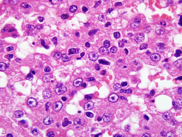
A large number of neoplastic lymphocytes had round or ovoid nuclei which were elongated or cleaved in some cells. The nuclei of neoplastic cells had darkly-stained coarse chromatin and were easily distinguishable from those of hepatocytes. The neoplastic cells had a scant amount of eosinophilic cytoplasm.
In addition, varying amounts of yellow pigment were engulfed in Kupffer cells. Neutrophils and other segmented nuclear granulocytes were increased in the sinusoids and emigrated into some hepatocytes and bile ducts. Immunohistochemically, large number of neoplastic cells showed positive reaction for CD3 (Fig. 2). Positive reaction for CD20, CD56, and CD79 was not observed. Immunopositive reaction for cleaved-caspase 3 was not observed in the hepatocytes with or without infiltrated neoplastic cells. Immunohistochemical examination for proliferating cell markers, such as Ki-67 and PCNA, brought equivocal results due to the intense positive reaction of infiltrating neoplastic cells in these hepatocytes.
Morphologic Diagnosis:
Lab Results:
Condition:
Contributor Comment:
The present case of feline malignant lymphoma involving the liver is considered to be the leukemic type due to the appearance of neoplastic cells in the sinusoids. Immunohistochemical findings suggest that the neoplastic lymphocytes are T-cell origin. According to the histologic criteria established by the World Health Organization (WHO), this case is T-cell chronic lymphocytic leukemia judging from the small cells with a dense chromatin distribution.
As stated above, neoplastic lymphocytes of T-cell origin occasionally have a character of infiltrating into the epidermis, the epithelium in adnexal tissues, or mucosal epithelium. In addition, as in the present case, the fact that CD3 positive neoplastic T-cell lymphocytes invaded the epithelium of a relatively large bile duct along with hepatocytes suggests that infiltration into hepatocytes reflects a common mechanism of neoplastic cells of T-cell origin.
In the present case hepatocellular damage due to intracellular invasion by neoplastic lymphocytes was considered possible; however, necrotic and apoptotic changes of hepatocytes were not detected morphologically and hepatocytes had a negative reaction for one of the enzymes concerning apoptosis, cleaved-caspase 3, by immunohistochemical examination. From these results, cytotoxic effect of infiltrating lymphocytes on hepatocytes was not evident.
JPC Diagnosis:
Conference Comment:
Humans have a distinct form of hepatic lymphoma termed sinusoidal T-cell lymphoma, and its histopathologic features share similarities with the case of this cat. The primary histologic finding in the human disease is diffuse infiltration of malignant T-cells into the hepatic sinusoids in the absence of a mass effect. The histologic observation of low to moderate numbers of sinusoidal lymphocytes in the liver biopsies and the clinical presentation of affected human patients often lead to the misdiagnosis of acute or chronic inflammatory liver disease.1 The sinusoidal form of hepatic malignant lymphoma in humans demonstrates the difficulty sometimes encountered when attempting to differentiate an inflammatory process from neoplasia by histopathology.
Among the various lymphoid neoplasms affecting cats, several features in this case are suggestive for large granular lymphocytic (LGL) lymphoma, including the contributors gross report of nodular lesions in the small intestinal, cytomorphology of neoplastic cells containing eosinophilic cytoplasmic granules, and prominent emperipolesis. The most common primary site for LGL lymphoma in the cat is the small intestine; additional information concerning the gross lesions found in the small intestine, including histopathologic findings, might have been useful in this case.
This case was studied in consultation with Dr. Peter Moore, a recognized expert in the field of veterinary hematopoietic neoplasia. Dr. Moore has observed a number of similar cases in which the neoplastic lymphoid cells in feline LGL lymphoma express CD3; based on this finding and other evidence, he suspects LGL type tumors arise from intestinal epithelial lymphocytes (IEL). Dr. Moore also has observed a similar hepatic infiltration pattern for LGL lymphoma in dogs, albeit without the intestinal association; publication of the canine form of the disease is forthcoming. An IEL origin for the malignant lymphoid cells might well explain the unique histologic infiltration pattern observed in the liver of this cat, as most cases of LGL lymphoma in cats have intestinal involvement with frequent involvement of the liver, and disseminating lymphomas frequently metastasize to the liver along hepatic sinusoids.(1,3)
We thank Dr. Moore for his informative consultation with this case.
References:
2. Ossent P, Stockli RM, Pospischil A: Emperipolesis of lymphoid neoplastic cells in feline hepatocytes. Vet Pathol 26:279-280, 1989
3. Valli VE: T-cell and NK-cell neoplasms. In: Veterinary Comparative Hematopathology, pp. 304-306. Blackwell Publishing, Ames, IA, 2007