Signalment:
Gross Description:
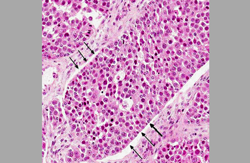
Histopathologic Description:
Immunohistochemically, the large round cells are positive for one of the germ cell markers, PLAP (placental alkaline phosphatase) and are negative for granulosa cell markers such as WT-1 and vimentin. In contrast, small spindle cells are positive for WT-1 and vimentin, and are negative for PLAP. Some tumor cells of middle-sized nuclei are positive for all three markers.
Morphologic Diagnosis:
Condition:
Contributor Comment:
The histological appearance of canine ovarian granulosa cell tumors is variable; testicular Sertoli cell tumor-like appearance is observed along with the follicular (i.e., micro- and macro- follicular), insular, diffuse, and trabecular growth patterns.(1,3) Granulosa cell tumors in the human ovary are further classified into adult and juvenile types. Juvenile granulosa cell tumors have solid and follicular growth patterns of tumor cells with abundant (luteinized) cytoplasm. Solid or follicular growth pattern of tumor cells with immature nuclei and abundant cytoplasm are thought to be important characteristics distinguishing juvenile from adult granulosa cell tumors.(8) The histological characteristic of the ovarian tumor in the present case is similar to the micro follicular pattern of canine granulosa cell tumors, but nuclear size varies among tumor cells and there are very large cells that appear similar to germ cells. According to the criteria for classification of human ovarian granular cell tumors, variation of nuclear size and the presence of very large nuclei, which may represent immature nuclei, suggest the diagnosis of juvenile granulosa cell. However, tumor cells characterized by large round nuclei and abundant cytoplasm in the present case are positive for PLAP (one of the germ cell markers), suggesting that these tumor cells are characteristic of germ cells rather than malignant neoplastic granulosa cells with immature nuclei. Although a majority of canine ovarian granulosa cell tumors are composed of multiple growth patterns in a single mass, the present tumor is composed of a single pattern throughout.(1,3) This growth pattern may be further evidence to rule out the diagnosis of granulosa cell tumor.Â
Patnaik and Mostofi first reported mixed germ cell-stromal tumors that were different from collision tumors of seminoma and Sertoli cell tumors in the testis of 16 dogs, whereas cellular components are identical in both tumors.(5) In their mixed germ cell-stromal tumors, the tumor is composed of uniform and close intermixing of two type cells: germ cells with a large round nucleus and Sertoli cells with a smaller elongated nucleus.(4) It is unlikely that the present tumor is a collision tumor of dysgerminoma and granulosa-theca cell tumor that originated in different sites of the ovary, because the histopathological features of the tumor were almost uniform throughout the mass.Â
Mixed germ cell sex cord-stromal tumors are further subdivided into two types: gonadoblastoma and mixed germ cell sex cord-stromal tumor. These two types of tumors are distinct clinicopathological and histopathological entities. In human cases, approximately 80 percent of gonadoblastomas occur in phenotypic female patients who have a Y chromosome (almost all have karyotypes of 46XY or 46XY/XO and dysgenetic gonads).(2,5,7) In contrast, mixed germ cell sex cord-stromal tumors develop in phenotypically and karyotypically normal females and males. Mixed germ cell sex cord-stromal tumors have been reported in male dogs with recognizable gonads.(2,4,5,7) Judging from normal development of the left ovary and other genital organs, the present dog was probably a genotypically normal female; however, an examination was not done on gene abnormality.
JPC Diagnosis:
Conference Comment:
The conference moderator led a practical discussion of immunohistochemical markers in human ovarian germ cell and sex cord-stromal tumors based on a recent report in Histopathology. The authors describe the use of SALL4 (Sal-like protein 4) and PLAP (placental alkaline phosphatase) to mark germ cell differentiation; OCT4 (octamer-binding transcription factor 4), CD117 and D2-40 to mark dysgerminoma; α-fetoprotein and glypican-3 to mark yolk sac tumors; OCT4, CD30 and SOX2 (sex determining region Y-box 2) to mark embryonal carcinoma; calretinin, inhibin, SF-1 (splicing factor 1) and FOXL2 (forkhead box L2) to mark sex cordstromal differentiation; and melan-A to mark steroid cell tumors.(6) The following is a summary of immunohistochemical stains used to differentiate human ovarian tumors as discussed in this article:
- PLAP is expressed in dysgerminomas, gonadoblastomas, embryonal carcoinomas, yolk sac tumors and choriocarcinomas.Â
- SALL4, a transcription factor required for development and maintenance of embryonic stem cell pluripotency, is more sensitive and specific than PLAP in identifying ovarian germ cell tumors; however, PLAP is more sensitive for choriocarcinomas.Â
- OCT4, a transcription factor required for maintaining embryonic stem cell pluripotency, is expressed in dysgerminomas, gonadoblastomas and embryonal carcinomas, whereas other germ cell tumors lack OCT4.Â
- CD-117 is a transmembrane tyrosine kinase growth factor receptor for stem cell factor (SCF) that shows strong membranous immunohistochemical staining in over 85% of dysgerminomas, and half of solid pattern yolk sac tumors. CD-117 is also overexpressed in other tumors, such as gastrointestinal stromal tumors and mast cell tumors; expression in these tumors is often cytoplasmic.Â
- D2-40, which marks the protein podoplanin, expressed in fetal germ cells, shows cytoplasmic and membranous expression in most dysgerminomas.Â
- NANOG is expressed in up to 83% of dysgerminomas and in the germ cell population of gonadoblastoma.Â
- SOX2 and CD30 expression is present in many embryonal carcinomas.Â
- Alpha-fetoprotein and glypican-3 is expressed in many yolk sac tumors.Â
- Glypican-3, a surface heparan sulphate proteoglycan that regulates cell growth during fetal development, is very specific for yolk sac tumors. Choriocarcinomas are also positive for glypican-3 but are usually easily differentiated from yolk sac tumors by histomorphology.Â
- HCG (human chorionic gonadotropin) is expressed by the syncytiotrophoblastic cells but not the mononuclear trophoblasts of an ovarian choriocarcinoma. Keratin, inhibin and glypican-3 are also expressed in choriocarcinomas.Â
- Inhibin is sensitive and specific for sex cord-stromal cell tumors.
- Calretinin, a maker of mesothelial differentiation, is more sensitive but less specific than inhibin for sex cord-stromal differentiation.Â
- SF-1, a transcription factor that regulates steroidogenesis, sexual differentiation and gonadal and adrenal gland development, is expressed in 100% of granulosa cell tumors.Â
- FOXL2, which encodes a transcription factor that is required for granulosa cell function and ovarian follicle development, is expressed in almost all granulosa cell tumors; it also appears to mark all types of sex cord-stromal tumors except for steroid cell tumors.Â
- WT-1 (Wilms tumor 1) is sensitive for sex cord-stromal differentiation.Â
- Steroid cell tumors express calretinin, inhibin, and SF-1and melan-A (also known as MART-1, or melanoma antigen recognized by T-cells 1).Â
- EMA (epithelial membrane antigen) is the best marker to exclude epithelial ovarian cancer.(6)
These markers have proven to be quite helpful in diagnosing ovarian germ cell tumors and sex cord-stromal tumors in humans; however, their efficacy in canine tumors has not been determined.Â
References:
2. Bolen JW. Mixed germ cell-sex cord stromal tumor. A gonadal tumor distinct from gonadoblastoma. Am J Clin Pathol. 1981;75:565-573.
3. Kennedy PC, Edwards JF, Goldschmidt MH, Larsen S, Munson L, Nielsen S. Histological classification of tumor of genital system of domestic animals. In: World Health Organization International Histological Classification of Tumors of Domestic Animals. Second Series. IV. Washington, DC: Armed Forces Institute of Pathology/ American Registry of Pathology; 1998.
4. Owston MA, Ramos-Vara JA. Histologic and immunohistochemical characterization of a testicular mixed germ cell sex cord-stromal tumor and a Leydig cell tumor in a dog. Vet Pathol. 2007;44:936-943.
5. Patnaik AK, Mostofi FK. A clinicopathologic, histologic, and immunohistochemical study of mixed germ cell-stromal tumors of the testis in 16 dogs. Vet Pathol. 1993;30:287-295.
6. Rabban JT, Zaloudek CJ. A practical approach to immunohistochemical diagnosis of ovarian germ cell tumours and sex cordstromal tumours. Histopathology. 2013;62: 7188.
7. Talerman A, Roth LM. Recent advances in the pathology and classification of gonadal neoplasms composed of germ cells and sex cord derivatives. Int J Gynecol Pathol. 2007;26:313-321.
8. Young RH. Sex cord-stromal tumors of the ovary and testis: their similarities and differences with consideration of selected problems. Mod Pathol. 2005;18 Suppl 2:S81-98.