Joint Pathology Center
Veterinary Pathology Services
Wednesday Slide Conference
2017-2018
Conference 4
September 20th, 2017
CASE I: N761-15 (JPC 4101761).
Signalment: 6-year-old, male, Criollo, Equus caballus, horse.
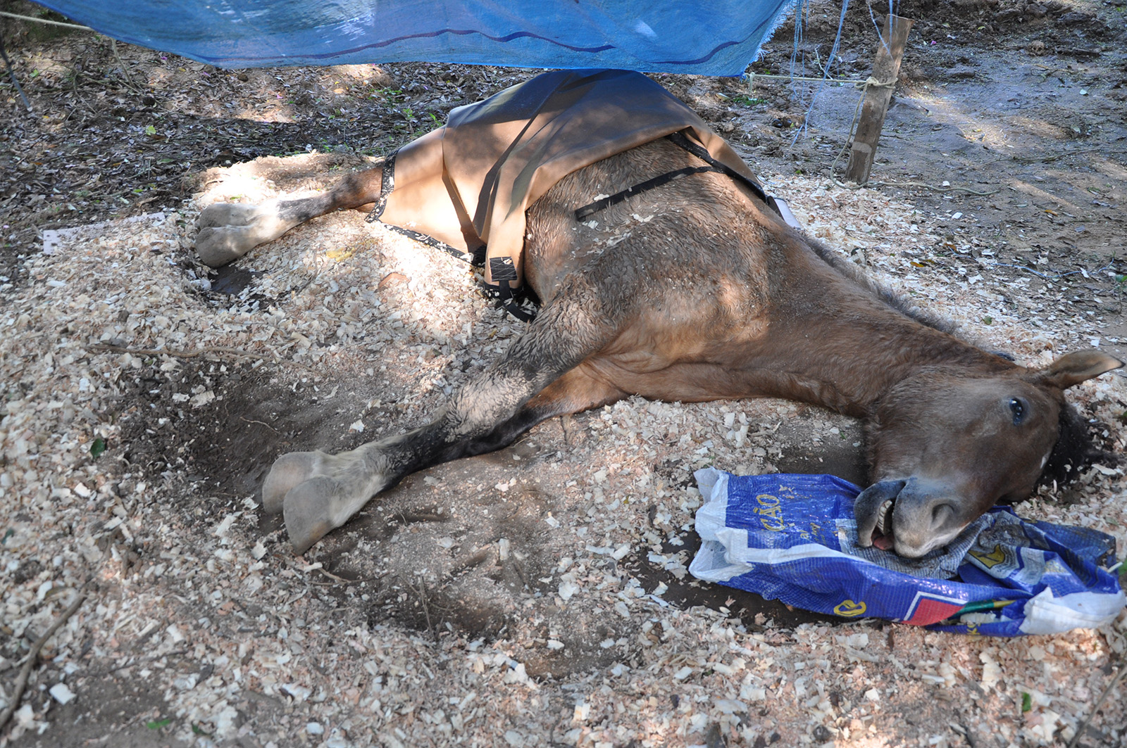
History: This horse lived in the metropolitan area of Porto Alegre since it was born. It had a nine day history of lethargy, sialorrhea, evolving to neurological signs of ataxia and recumbency. Euthanasia was elected due to the poor prognosis and progression of clinical signs. Two days prior to onset of clinical signs, Trema micrantha branches were pruned, and its leaves were readily available for consumption in the pasture the horse was hold.
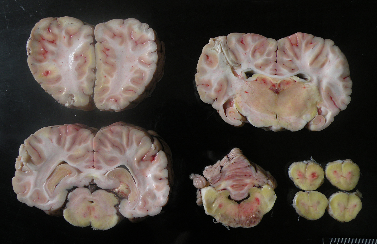
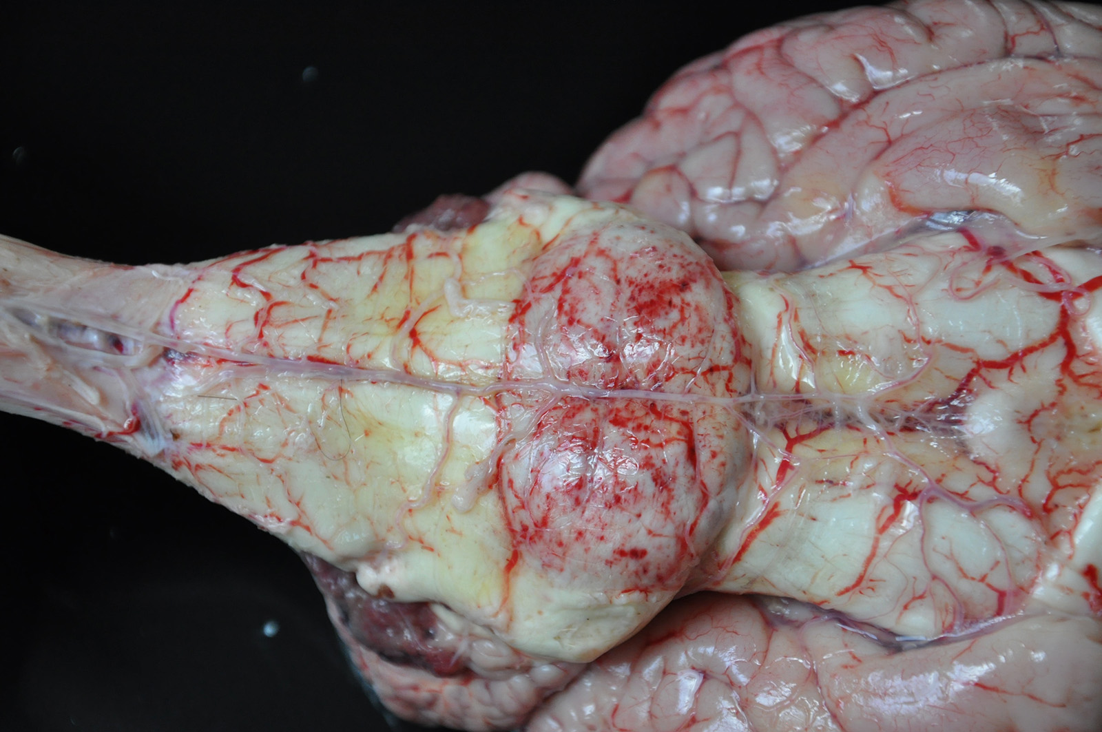
Gross Pathology: The brain of this horse was diffusely yellow with grayish to dark-red multifocal do coalescent friable to soft pinpoint foci, mainly involving the pons.
Laboratory results: Fragments of cerebrum, cerebellum and spinal cord were refrigerated and tested by direct fluorescent antibody test (DFAT) for Rabies virus (RABV) in a certified laboratory, following OIE recommendations, and gave negative results.
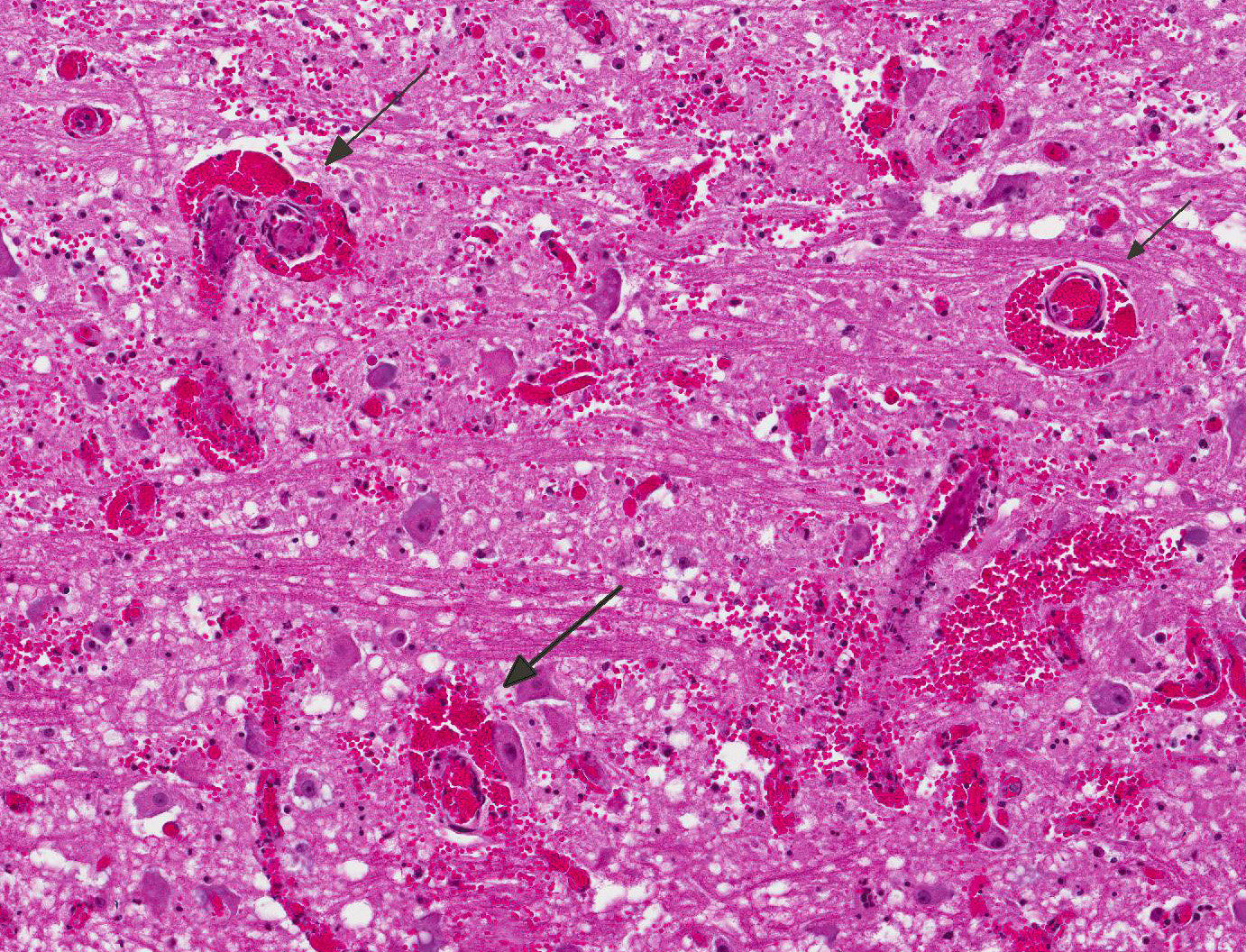
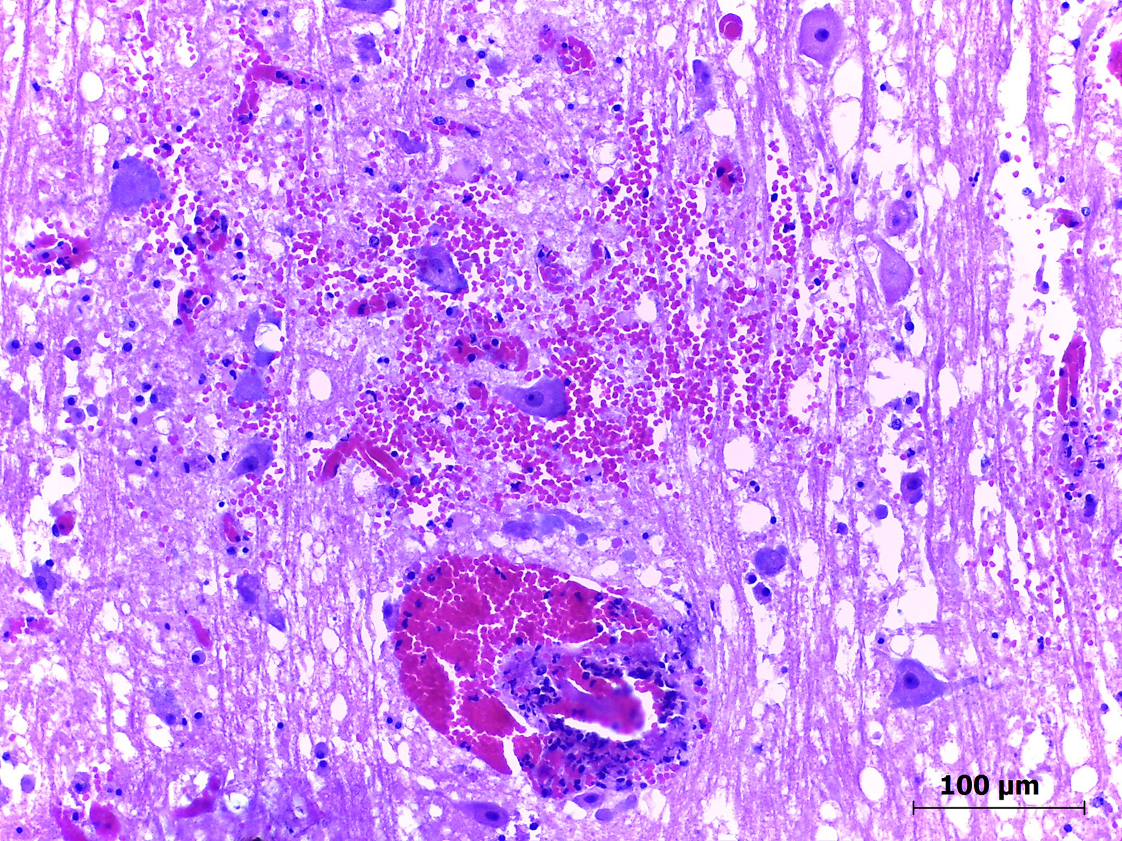
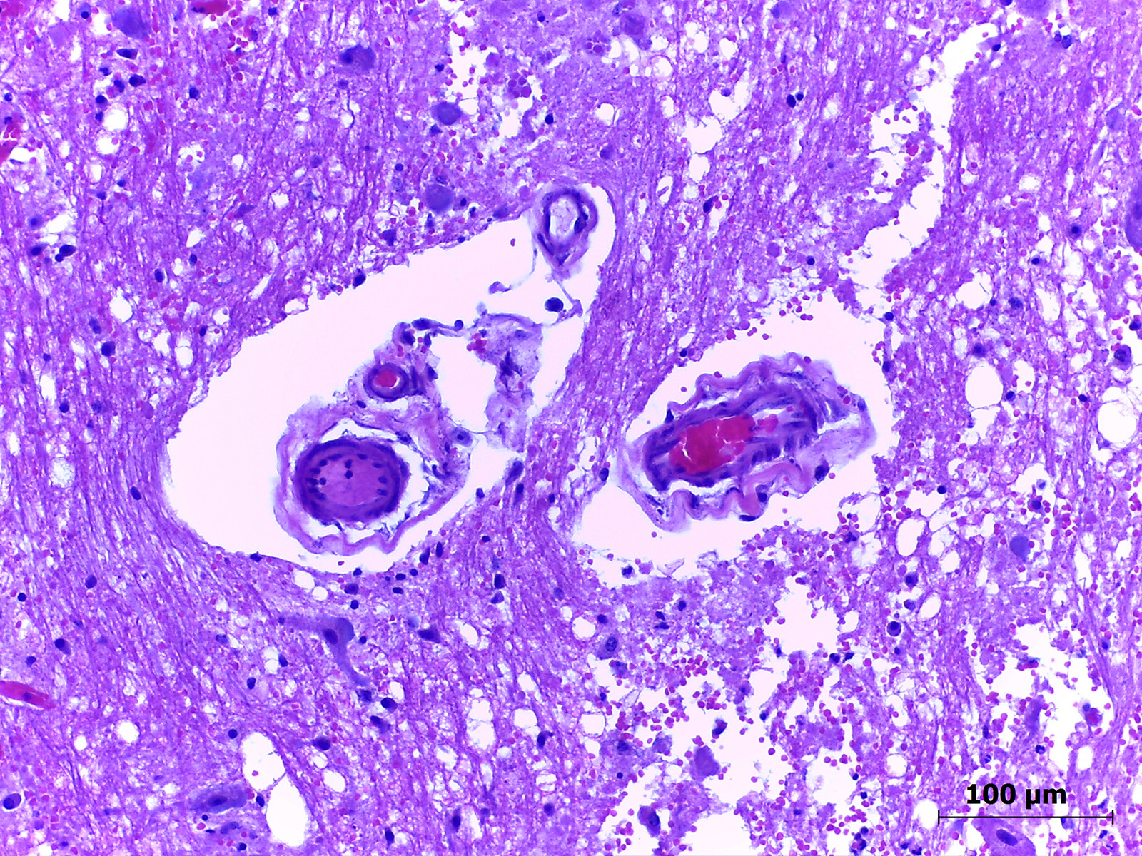
Microscopic Description: Pons: Approximately one third of the cut section (which corresponds to the pons) presents severe vasculitis and liquefactive necrosis of white and gray matter; these are characterized by multifocal transmural fibrinoid necrosis of blood vessels, which sometimes are occluded by thrombosis and associated with perivascular hemorrhage, in addition to severe vacuolation of myelin (suggestive of intramyelinic edema) and perivascular edema. There is a moderate multifocal perivascular inflammatory infiltrate consisting predominantly of neutrophils with few lymphocytes and plasma cells. At the periphery of the areas with necrosis of blood vessels, numerous Gitter cells, Wallerian degeneration and multiple axonal spheroids are observed.
Contributors Morphologic Diagnosis: Brain (pons): Focally extensive, liquefactive necrosis of the white and gray matter, with severe multifocal vasculitis, fibrinoid necrosis, thrombosis, perivascular hemorrhage and intramyelinic edema.
Condition: Encephalomalacia due to Trema micrantha poisoning
Contributors Comment: This horse was part of a major study involving 14 horses that were poisoned by T. micrantha consumption in different municipalities of Rio Grande do Sul state, Brazil. This condition caused lesions that affected different regions of the CNS, but the most striking lesions were observed in pons.
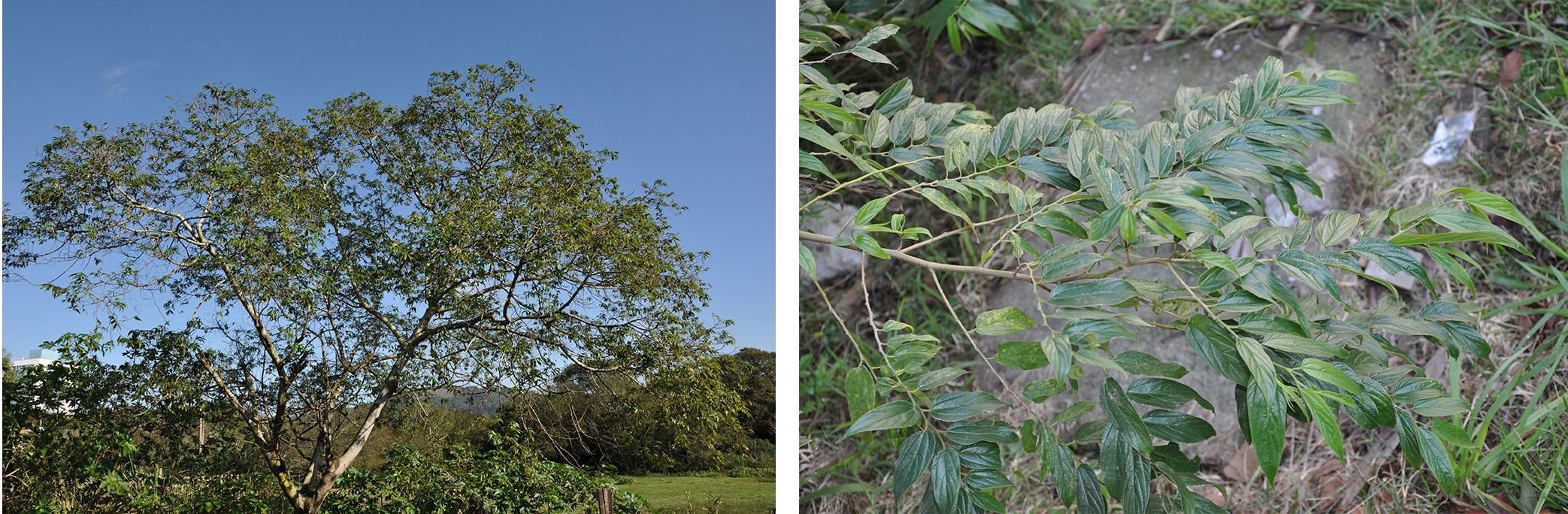
T. micrantha is an arboreal species widely distributed in Brazil, within the Cannabaceae family, occurring in tropical and subtropical areas in almost all tropical and subtropical areas of South, and Central America countries4, and in the southern counties of Florida (United States of America).4 It is a fast growing tree up to 5-20m, that has highly palatable leaves, which are promptly consumed by herbivores, especially when branches with green leaves fall to the ground, either due to pruning or windstorms, becoming readily available for consumption,9 as in the present case.
The toxic compounds of T. micrantha are still unknown,8 though a toxic compound named trematoxin obtained from Trema tomentosa has been described in Australia. This is a hepatotoxic glycoside associated with centrilobular necrosis in cattle, sheep, goats, horses and camels;10 however hepatic lesions were absent in this horse, as in the majority of the remaining.
Neurological abnormalities after T. micrantha consumption have been related to hepatic encephalopathy, resulting in Alzheimer type II astrocytes and perivascular edema on histopathology of the CNS from the affected animals1. However, lesions of this horse were mainly characterized by malacia, severe fibrinoid degeneration of blood vessels, thrombosis and hemorrhage. The massive necrosis and softening of the brain may be attributed to an ischemic lesion secondary to the fibrinoid necrosis of blood vessels, which resulted in extensive areas of hemorrhage and thrombosis and, consequently, areas of liquefactive necrosis, as previously described. These are not lesions usually related to hepatic encephalopathy. Still, the cause of these lesions is unknown, however it is speculated that it results from the action of an intermediary metabolite formed immediately after T. micrantha consumption, which would only occur in equid metabolism.6
Differential diagnosis of the present case included parasitic infection by Trypanosoma evansi and leukoencephalomalacia due to the prolonged ingestion of corn contaminated with fumonisin B1.7 This horse did not have access to contaminated corn, and lesions differed from that condition due to the involvement of both gray and white matter. T. evansi infection in horses is characterized by a non-suppurative encephalitis and edema,7 which was not observed in this horse.
In this case, T. micrantha consumption caused predominantly a neurological disease, with absent hepatic lesions. Thus, this neurotoxicosis should be considered in the differential diagnosis of CNS diseases in horses.
JPC Diagnosis: Brain, pons: Vasculitis, necrotizing, multifocal, marked with thrombosis, edema, and perivascular hemorrhage, Criollo, equine.
Conference Comment: This case is an unusual presentation of toxic encephalomalacia caused by Trema micrantha ingestion. The contributor provided an excellent review of Trema micrantha and T. tomentosa poisoning which was mirrored in much of the conference discussion. Conference participants reviewed several differentials, including gram-negative sepsis, purpura hemorrhagica, equine herpesvirus-1, listeriosis, eastern equine encephalitis (EEE), leukoencephalomalacia, Trypanosoma evansii and cerebrovascular accidents. Most differentials could be ruled out based on lesion distribution and host inflammatory response. Equine herpesvirus-1 is most common in the white matter of the spinal cord and characteristically results in nonsuppurative necrotizing vasculitis and thrombosis.2 Listeria monocytogenes, is common in the brainstem, but is characterized by microabscesses composed of small aggregates of neutrophils within the neuroparenchyma.2 The equine encephalitides (Alphaviruses) produce diffuse lesions in the grey matter with increasing severity in the cerebral cortex consisting of lymphoplasmacytic and neutrophilic necrotizing encephalitis.2
The differentials discussed in-depth include leukoencephalomalacia, purpura hemorrhagica, and cerebrovascular accident, because they are associated with encephalomalacia without significant inflammation of the neuroparenchyma, as is seen in this case.
Leukoencephalomalacia (LEM), also known as moldy corn poisoning, occurs in horses, donkeys, or mules fed corn laced with mycotoxin fumonisin B1 which is produced by Fusarium verticillioides or F. proliferatum, which grow in warm, moist conditions causing sporadic outbreaks of disease. Fumonisin causes encephalomalacia of the white matter in two ways (1) it damages the microcirculatory system and impairs cardiovascular function, and (2) competitively inhibits sphingosine N-acetyltransferase which leads to the accumulation of sphingosine and blocks the production of spingolipids. In this case, lesions were in both the grey and white matter of the pons. In contrast, LEM produces lesions in the white matter of the cerebrum.2, 5
Although often associated with myositis, purpura hemorrhagica can affect many organs as it results in tissue necrosis secondary to vascular injury caused by immune-complex deposition (IgA and streptococcal M protein). In horses, purpura hemorrhagica accompanies Streptococcus equi infection and results in fibrinonecrotic vasculitis and hemorrhagic infarcts. Inflammatory infiltrates are rarely seen, but when seen are usually located at the periphery of areas of necrosis and Streptococcus equi is isolated from the lymph nodes or guttural pouch.3
Cerebrovascular accidents (CVA) which are increasing in recognition in small animals due to increasing awareness of their existence as well as sophisticated imaging techniques are often characterized by ring hemorrhage in the absence of significant inflammation. Literature on this problem in large animals is currently lacking.
Faculdade de Veterinária
Universidade Federal do Rio Grande do Sul
Setor de Patologia Veterinária
http://www.ufrgs.br/patologia/
References:
1. Bandarra PM, Pavarini SP, Raymundo DL, et al. Trema micrantha toxicity in horses in Brazil. EquineVet J. 2010;42:456-459.
2. Cantile C, Youssef S. Nervous system. In: Maxie MG, ed. Jubb, Kennedy, and Palmers Pathology of Domestic Animals. Vol. 1. 6th ed. St. Louis, MO: Elsevier; 2016:302, 315-316, 362-364, 376-377, 383-384.
3. Cooper BJ, Valentine BA. Muscle and tendon. In: Maxie MG, ed. Jubb, Kennedy, and Palmers Pathology of Domestic Animals. Vol. 1. 6th ed. St. Louis, MO: Elsevier; 2016:229.
4. Hargis AM, Myers S. The integument. In: Zachary JF ed. Pathologic Basis of Veterinary Disease. 6th ed. St. Louis, MO: Elsevier; 2017:1120-1121.
5. Lorenzi H. Árvores Brasileiras. Manual de Identificação e Cultivo de Plantas Arbóreas Nativas do Brasil (Handbook for identification and cultivation of native trees from Brazil).5th ed. São Paulo, Brazil: Plantarum; 2008;01:90.(In Portuguese)
6. Miller AD, Zachary JF. Nervous system. In: Zachary JF ed. Pathologic Basis of Veterinary Disease. 6th ed. St. Louis, MO: Elsevier; 2017:879-880.
7. Nelson G. Elms and mulberries. In: The trees of Florida: a reference and field guide. 1st ed.: Pineapple Press Inc., Sarasota. Florida. 1994:48-52.
8. Pavarini SP, Bandinelli MB, Bassuino DM, et al. Novos aspectos sobre a intoxicação por Trema micrantha (Cannabaceae) em equídeos. Pesq Vet Bras. 2013;11:1339-1344.
9. Rech RR, Barros CSL. Neurologic diseases in horses. Vet Clin North Am EqPract. 2015;31:281-306.
10. Tokarnia CH, Brito MF, Barbosa JD, et al. Plantas Tóxicas do Brasil (Poisonous plants from Brazil). 2nd ed. Rio de Janeiro, Brazil: Helianthus; 2012:174-176. (In Portuguese)
11. Traverso SD, Corrêa AMR, Schmitz M, et al. Intoxicação experimental por Trema micrantha (Ulmaceae) em bovinos. Pesq Vet Bras. 2004;24:211-216.
12. Trueman KF, Powell MV. Suspected poisoning of camels by Trema tomentosa (poison peach). Aust Vet J. 1991;68:213-214.