Signalment:
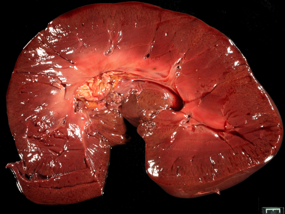
Gross Description:
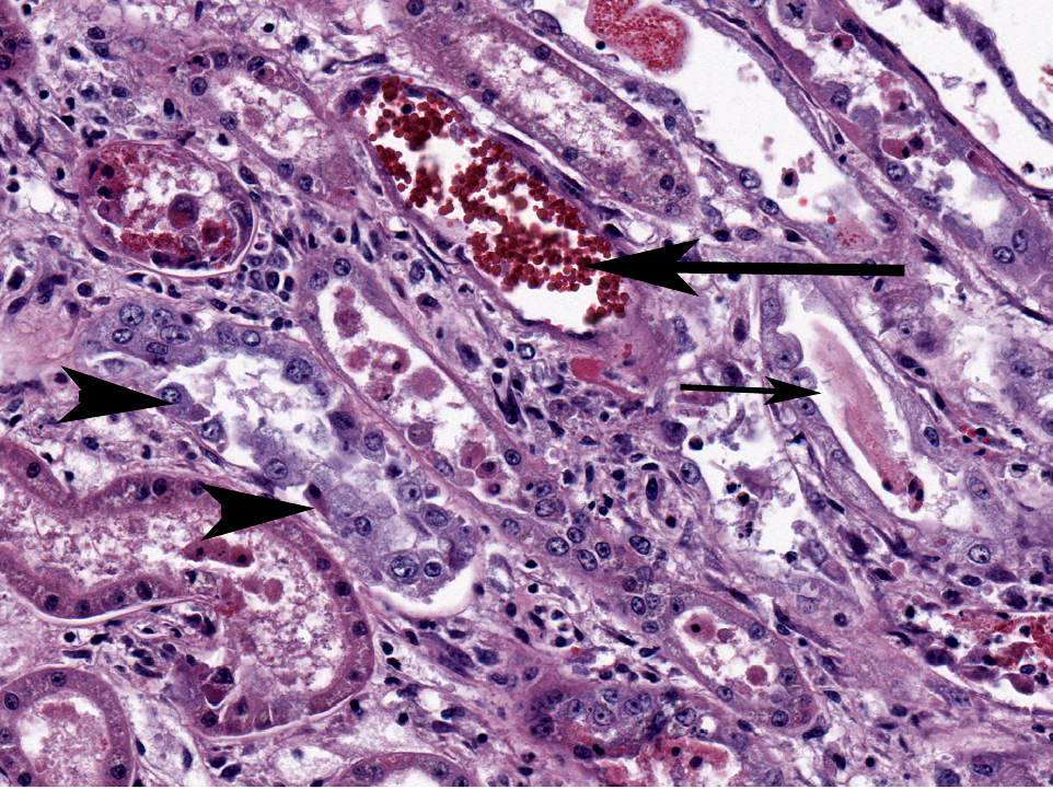
Histopathologic Description:
Morphologic Diagnosis:
Lab Results:
| Hemogram |
| Leucocytosis with absolute mature neutrophilia (28975 {2260-8580}) |
| Mild absolute monocytosis |
| Mild lymphoctosis |
| 2+ eccentrocytes |
| Serum = 4+ hemolysis |
| Creatinine kinase 4683U/L (73-450)* |
| Aspartate aminotransferase 2246U/L (134-643)* |
| Total protein 11.6 g/dl (5.3-7.3)* |
| BUN 58mg/dl (7-28) |
| Creatinine 4.3mg/dl (1.1-2) |
| Total bilirubin 10.3mg/dl (<4.1)* |
| * tests can be falsely elevated when hemolysis >2+ |
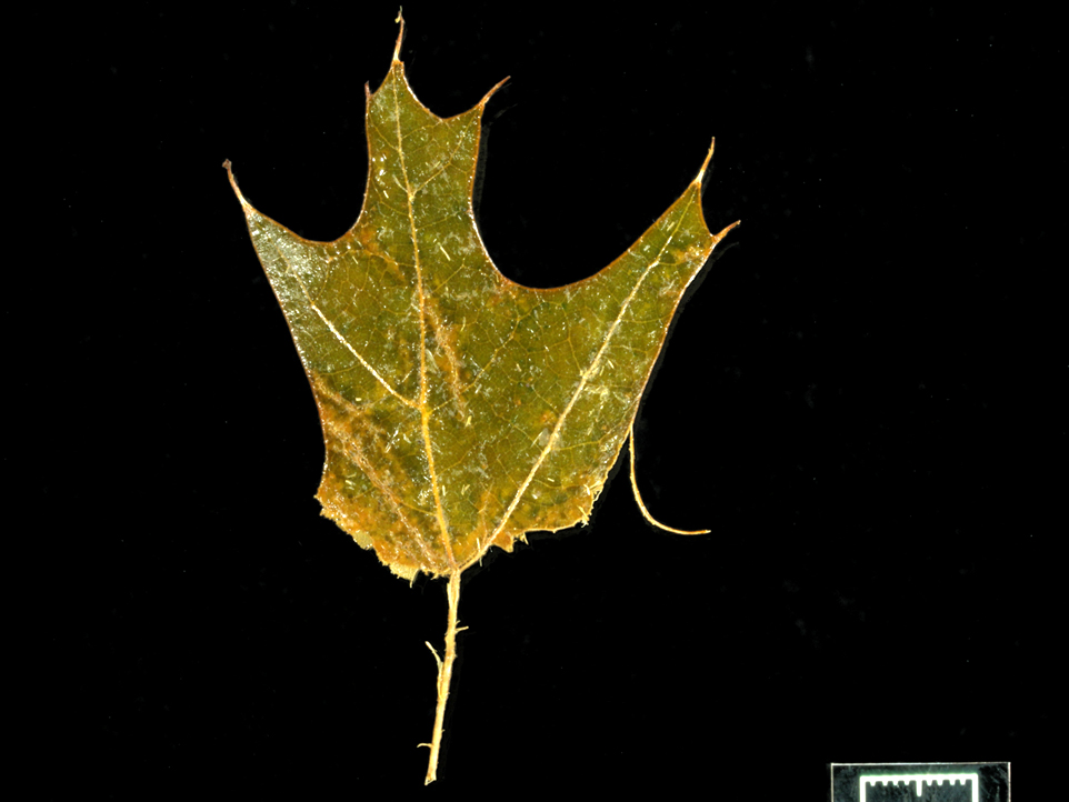
Condition: Red maple leaf toxicity
Contributor Comment:
- the kidney was/is remarkably preserved despite autolysis in other tissue
- note where the iron metal stains
- it is a segmental nephrosis
- the karyomegaly often is not emphasized.(6)
The toxic principle is in wilted leaves, and its identity is unknown. Gallic acid is one of the usual suspects as an oxidant, and Vitamin C (and steroids) is recommended for treatment.(1,8) The combination of intravascular hemolysis, hemoglobinemia and hypoxemia make the renal lesion. Reasonable differential diagnoses include: onion toxicity (we have cases of wild onion toxicity in horses but it is milder), nitrite intoxication (iatrogenic), equine infectious anemia (fever), babesiosis (fever and parasites visible) and immune mediated hemolytic anemia (agglutination positive). We have had cases associated with silver maple.
JPC Diagnosis:
Conference Comment:
There are two forms of A. rub rum toxicosis: a peracute form that typically occurs late in the autumn and a hemolytic form which occurs in early autumn. The peracute form results in massive methemoglobinemia, tissue anoxia and sudden death. Gross lesions include brown discoloration of tissues and blood, and cyanotic mucous membranes. The hemolytic form causes Heinz body formation, and subsequent intra- and extravascular hemolysis, in combination with methemoglobinemia. Clinical signs include weakness, lethargy, depression, anemia, icterus, hemoglobinuria and hemoglobinemia. Gross lesions include icterus, serosal petechia and ecchymosis, brown discoloration of the kidneys, splenomegaly, pulmonary congestion and edema, and mild to severe centrilobular hepatic degeneration and lipidosis.(5,7,13)
Differential diagnoses for hemolytic anemia in horses include Leptospira bratislava, Babesia caballi and B. equip, and wild onion (Allium sp.), which cause intravascular hemolysis; equine infectious anemia and Anaplasma plasmaphagocytophilum cause extravascular hemolysis. Other causes of methemoglobinemia include nitrate poisoning, chlorate toxicosis, and drugs such as phenacetin and acetanilide. Additional causes of Heinz body anemia include onion consumption in cattle, horses, dogs, and cats; rape, kale, and turnip consumption in cattle and sheep; chronic copper toxicosis in sheep, cattle, and swine; zinc toxicity in dogs; selenium deficiency; and the urinary acidifier methylene blue in cats. Heinz bodies can also occur spontaneously in cats.(5,7,13)
References:
2. Brockus CW. Erythrocytes. In: Latimer KS, ed. Duncan & Prasses Veterinary Laboratory Medicine Clinical Pathology, 5th ed. Ames, IA: John Wiley & Sons Inc; 2011:34-5.
3. Corriher CA, Gibbons DS, Parviainen, AKJ, et al. Compend Cont Educ Prac Veter. 1999;21:74-80.
4. Divers TJ, George LW, George JW. Hemolytic anemia in horses after the ingestion of Red Maple leaves. JAVMA. 1982;180:300-302.
5. Fry MM, McGavin MD. Bone marrow, blood cells, and lymphatic system. In: McGavin MD, Zachary JF, eds. Pathologic Basis of Veterinary Disease.5th ed. St. Louis, MO: Mosby Elsevier; 2011:718.
6. George LW, Divers TJ, Mahaffey EA, et al. Heinz body anemia and methemoglobinemia in ponies given Red Maple (Acer rubrum L.) leaves. Vet Pathol. 1982;19:521-533.
7. Maxie MG, Newman SJ. Urinary system. In: Maxie MG, ed. Jubb, Kennedy, and Palmers Pathology of Domestic Animals. 5th ed. St. Louis, MO: Saunders Elsevier; 2007:446-447.
8. McConnico RS, Brownie CF. The use of ascorbic acid in the treatment of 2 cases of Red Maple (Acer rubrum)-poisoned horses. Cornell Vet. 1982;82:293-300.
9. Plumlee KH. Red Maple toxicity in a horse. Vet Hum Toxicol. 1992;33:66-67.
10. Semrad SD. Acute hemolytic anemia from ingestion of Red Maple leaves. Compend Cont Educ Pract Veter. 1993;15:2, 261-264.
11. Stair EL, Edwards WC, Burrows GE, et al. Suspected Red Maple (Acer rubrum) Toxicosis with Abortion in Two Percheron Mares. Vet Hum Toxicol. 1993;35:229-230.
12. Tennant B, Dill SG, Glickman LT, et al. Acute Hemolytic Anemia, Methemoglobinemia, and Heinz Body Formation Associated with Ingestion of Red Maple leaves by Horses. JAVMA. 1981;179:143-150.
13. Valli VEO. The hematopoietic system. In: Maxie MG, ed. Jubb, Kennedy, and Palmers Pathology of Domestic Animals. 5th ed. St. Louis, MO: Saunders Elsevier; 2007:254-255.
14. Warner AF. Methemoglobinemia and hemolytic anemia in a horse with acute renal failure. Compend Cont Educ Pract Veter. 1984;6:S465-8, S472.