Signalment:
Gross Description:
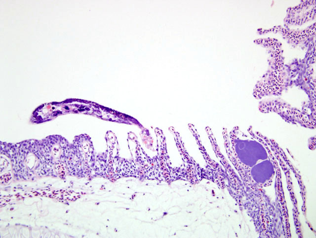
Histopathologic Description:
Morphologic Diagnosis:
Lab Results:
Condition:
Contributor Comment:
Freshwater fish infected with heavy gill infestations result in respiratory disease. Clinical signs can include opaque mucus covering the gills, protrusion of the gill filaments from under the gill covers and gills may be swollen and pale. Infected fish are less tolerant of low oxygen conditions and have an increased respiratory rate with gulping of air at the water surface. Fish become anoxic with flaring of the gill opercula.1 At a very advanced stage, the fish will isolate itself and spend long periods lying on the bottom with its fins clamped to its body. Acute infections are characterized by a short period of dyspnoea followed by sudden death.1
Most monogenean flukes are browsers, moving about the body surface and feeding on dermal mucus and gill debris. Monogeneans have a series of hooks that enable them to attach while feeding.2 Flukes anchoring to the gills induce a variety of lesions, depending on the density and species of parasite. Lesions can range from excessive mucous secretions, hyperplasia of gill epithelium with fusion of secondary lamellae to the presence of haemorrhage, necrosis / ulceration and inflammation.3,6 Secondary infection by bacteria and fungi are commonly established at damaged epithelial sites.4
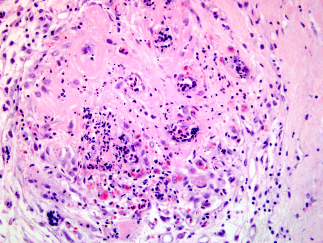
In this case, there is a widespread arterial vasculitis, which we propose may be mediated by immune complexes. Other discus fish in the collection are almost certainly affected by flukes but the involvement of a vasculitis, in other cases, remains to be determined.
The Dactylogyrus gill fluke life cycle is direct, not requiring intermediate hosts. The adults are oviparous and produce eggs with long filaments. The eggs are usually attached to the gills and develop into a free-swimming onchomiricidium, which then locates and attaches to the fish within a few hours.5
JPC Diagnosis:
2. Gill: Vasculitis, necrotizing, multifocal, moderate with edema and hemorrhage (Fig. 1-2).
Conference Comment:
Conference participants noted a microsporidian-like organism within some of the sections examined. Coinfections are not uncommon within compromised gill epithelium.7
Other diseases of importance that may affect the gills include the following list adapted from Moeller4 and Wootten8
- Bacterial
- Flexibacter columnaris, Flexibacter psychrophilus, Cytophaga psychrophilia, and Flavobacterium - prominent hyperplasia, clubbing adn fusion of lamella, necrosis of gill lamella
- Fungal
- Branchiomyces sanguinis and B. demigrans (gill rot) - fungal disease of carp, trout and eels, prominent gill necrosis, with hyphae
- Saprolegnia, Achyla, Aphanomyces (Saprolegniasis) - white to brown cotton-like growths on skin, fins, and gills
- Protozoal
- Ichthyophthirius multifiliis (Ich) - ciliated protozoan with horseshoe nucleus, hyperplasia with encysted trophozoites on skin and gills
- Aurantiactinomyxo sp (myxosporidean) - hamburger gill disease, granulomatous inflammation and swelling of gills, with epithelial hyperplasia and gill necrosis surrounding cysts
- Microsporidians (Glugea, Pleistophora, Loma) - cysts contain 1-2 um spores
- Trematode
- Diplostomum spathaceum - (eye fluke) - digenetic trematode, gulls/pelicans definitive host, metacercaria in the anterior chamber, vitreous body, and lens, snails 1st intermediate host, salmonids 2nd intermediate host
- Gyrodactylus sp. - a monogenetic trematode that attaches to skin, fins, and gills
- Uvulifer ambloplitis (black spot disease) - digenetic fluke, numerous black to brown spots over skin, gills, and eyes, snails 1st intermediate host, fish 2nd intermediate host
- Other
- Argulus sp. - (fish louse) parasite of the skin and buccal cavity resulting in cutaneous ulcers, contains a retractile preoral stylet used to pierce the skin
- Lernea sp. (at base of fins) & Ergasilus sp. (on the gills) - Copepod, invades the skin, forms ulcers that are slow to heal
- Cryptosporidiosis - intracellular extracytoplasmic protozoan,
References:
2. Ferguson HW: Gills and pseudobranchs. In: Systematic Pathology of Fish, eds. Herman RL, Meade JW, pp. 11-40. Iowa State University Press, Ames, IA, 1989
3. Herbert BW, Shaharom FM, Anderson IG: Histopathology of cultured sea bass (Lates calcarifer) (Centropomidae) infected with Cruoricola lates (Trematoda: Sanguinicolidae) from Pulau Ketam, Malaysia. Int J Parasitol 25:3-13, 1995
4. Moeller RB: Diseases of Fish. found at http://www.afip.org/consultations/vetpath/moeller01.pdf, 2001
5. Roberts RJ: The pathophysiology and systemic pathology of teleosts. In: Fish Pathology, ed. Roberts RJ, pp. 55-132. WB Saunders, London, 2001
6. Roubal FR: Microhabitats, attachment of eggs and histopathology by the monogenean Allomurraytrema robustum on Acanthopagrus australis (Pisces: Sparidae). Int J Parasitol 25:293-298, 1995
7. Smith SA, Noga EJ: General parasitology. In: Fish Medicine, ed. Stoskopf MK, pp. 132-148. W.B. Saunders Company, Philadelphia, PA, 1993
8. Wooten R: The parasitology of teleosts. In: Fish Pathology, ed. Roberts RJ, pp. 242-288. WB Saunders, London, 2001