Signalment:
Gross Description:
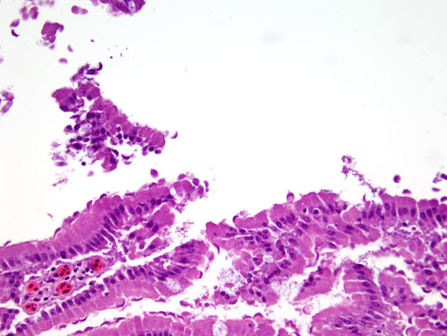
Histopathologic Description:
Morphologic Diagnosis:
Lab Results:
Condition:
Contributor Comment:
Giardia colonize the upper small intestine, usually the duodenum, in the trophozoite form.(1) The trophozoites are piriform, with four paired flagella, paired nuclei and a ventral disk on the concave surface which attaches to the host enterocyte. Reproduction by binary fission occurs in the gut lumen. This form may be passed in the feces, but more commonly, oval cysts are seen in direct smears. Transmission of the environmentally-stable cyst is by the fecal-oral route.(1) Diagnosis is best made by seeing the organism in a direct fecal preparation from a clinically ill animal, but ELISA and PCR tests have been developed.
Though giardiaisis is generally an asymptomatic condition, the presence of the organism has been associated with vitamin E/selenium deficiencies in cockatiels.(6) The proposed mechanism involves malabsorption of these nutrients due to the presence of the organisms on the intestinal mucosal surface. Though speculative, hypophosphatemia in this white-faced ibis may also have resulted from a focally extensive and heavy parasite load. Phosphorus is absorbed from the diet in the proximal small intestine where the heaviest load of parasitism was found in this bird. The bird of this report had adequate fat and muscling, but antemortem bloodwork revealed a PO4 of 0.7mg/dl (ref. 3.1-6.6mg/dl). Additionally, marked thoracolumbar scoliosis was present in addition to the fractured and dislocated digits. Thus, while the protist in this particular animal did not appear to cause a direct effect on intestinal health, it may have acted indirectly on bone health by blocking absorption of some nutrients needed for maintenance of healthy, structural bone.
JPC Diagnosis:
Conference Comment:
Giardia is an ubiquitous organism with worldwide distribution and is the most common flagellate of mammals and birds(3). Giardiasis is also a zoonotic disease. The life cycle of Giardia species is direct. Trophozoites and cysts are present in feces and passed from infected hosts into the environment. Once outside the host the trophozoites die, but the cysts are resistant. They are protected from the environment by a membranous layer with an inner and outer cyst membrane and filamentous layer forming a dense mat of interlacing branches.(2) Cysts are ingested via fecal-oral transmission. Enzymes within the stomach and small intestine cause excystation releasing two trophozoites. The organisms attach and feed in the upper small intestine and multiply by binary fission. Once the organisms reach the colon, they encyst in preparation for the external environment.Â
References:
2. Cheville NF: Ultrastructural Pathology, pp. 722-723. Iowa State University Press, Ames, Iowa, 1994
3. Gardiner CH, Poynton SL: An Atlas of Metazoan Parasites in Animal Tissues, 2nd ed., pp. 6-7. Armed Forces Institute of Pathology, Washington, DC 1984
4. McRoberts KM, BP Meloni, UM Morgan, R Marano, N Binz, SL Erlandsen, SA Halse, RCA Thompson: Morphological and molecular characterization of Giardia isolated from the straw-necked ibis. J. Parasitol. 82(5):711-718, 1996
5. Monis PT, RCA Thompson: Cryptosporidium and Giardia-zoonoses: fact or fiction? Infection, Genetics and Evolution 3:233-244, 2003
6. Ritchie B, GJ Harrison, LR Harrison: Avian Medicine: Principles and Application. pp. 732, 1014-5. Wingers Publishing, Lake Worth, FL, 1994