Signalment:
38 week old male Sprague Dawley rat,
Rattus norvegicusThe rat was euthanized at week 38 of a chronic toxicity study due to a large mass involving the left maxillary region.
Gross Description:
A red, firm mass was noted that involved the entire left maxillary region. It was 35 X 25 X 20 mm. The mass appeared the same on the cut surface.
Histopathologic Description:
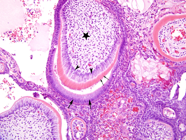
The normal bony architecture of the maxilla is largely replaced by a poorly circumscribed
mass (4-1) that is predominantly composed of epithelial and mesenchymal elements that frequently form variably-sized and shaped tooth-like structures. The epithelial elements of the mass include ameloblasts and odontoblasts that are generally well-differentiated; these cells often palisade along eosinophilic extracellular material with fine, tubular cavities (dentin) or lesser amounts of a more basophilic, hyaline material (enamel). Spindled to stellate mesenchymal cells with an accompanying vascular component (dental pulp) are often present centrally within the abortive tooth-like structures. Multifocally within the mass, odontogenic cells occasionally form cords and nests that are not associated with dental hard substance; these structures sometimes have central cavitations that contain degenerating cells and necrotic debris. There are multifocal areas of necrosis, inflammation, hemorrhage, pigment, and new bone formation within or at the periphery of the mass.
Morphologic Diagnosis:
Odontoma
Condition:
Compound odontoma
Contributor Comment:
An odontoma is a dental neoplasm (or hamartoma) in which cellular maturation and differentiation have progressed to the stage of development of both enamel and dentin. Two types are recognized: complex odontoma and compound odontoma. Complex odontomas contain dental pulp, mesenchymal cells, and hard tissue elements, but there is poor differentiation of the cellular components such that the mass has little resemblance to normal tooth architecture. Compound odontomas have a higher degree of cellular differentiation, and their hard tissue elements resemble abnormally shaped tooth-like structures (denticles). The hard tissue generally appears as dentin (an acellular, smooth, eosinophilic material) with smaller amounts of cementum (resembling bone). Enamel may be completely removed upon decalcification, in which case it will appear as a clear space or cleft in close apposition to the dentin. Incompletely decalcified specimens may have a small amount of enamel that stains basophilic with hematoxylin and eosin.
9 Complex odontomas are rare in all species but are most commonly seen in young horses and young dogs. Compound odontomas generally present as mass-like lesions of the jaw of young canines.
7 Both types of odontomas have been described in the rat.
3, 9 In those species such as the rat where incisors grow continuously throughout the animals lifetime, odontoma must be distinguished from dysplasia or a malformation that is congenital or secondary to malocclusion or fracture of the incisor tooth.
3
Odontomas and other odontogenic tumors have been experimentally induced in rats by carcinogens such as methylnitrosourea and related compounds.
4
JPC Diagnosis:
Bone, maxilla: Compound odontoma, Sprague-Dawley rat (Rattus norvegicus), rodent.
Conference Comment:
Currently there is controversy on the classification of odontomas as either neoplasms
7 or non-neoplastic hamartomas.
6 Odontomas are tumors in which there is a combination of both odontogenic epithelial components and dental matrix structures such as dentin and enamel. The inductive theory of odontogenesis states that the ameloblastic epithelium promotes the surrounding mesenchymal cells to become odontoblasts. These odontoblasts produce dentin, which is necessary for the ameloblasts to form enamel. Neoplasms composed of only epithelium without hard tissues are termed ameloblastomas.
8
Odontomas can be classified into various types based on their components and organization:
- Complex odontoma2 Contains well differentiated dental tissues, including dentin, enamel matrix, odontogenic epithelium, and cementum (horses and rodents) that do not form tooth-like structures
- Compound odontoma2 Contains cords of odontogenic epithelium, with intermittent complete odontogenesis forming tooth-like structures (denticles). Occasionally there is bone matrix formation surrounding or adjacent to the denticles.Â
- Odontoameloblastoma2 Contain areas of ameloblastic epithelium that are separate from other areas of complex or compound odontomas.
- Ameloblastic fibro-odontoma2 Contain both dental epithelial tissues (resembling dental lamina) and mesenchymal tissues (resembling dental pulp) that are associated with enamel and dentin.Â
- Dentinoma3 Contain odontoblasts producing a calcified dentin tissue with no evidence of enamel formation.
References:
1. Boy SC, Steenkamp G: Odontoma-like tumours of squirrel elodont incisors-elodontomas. J Comp Pathol 135:56-61, 2006
2. Brown CC, Baker DC, Barker IK: Alimentary system. In: Jubb, Kennedy, and Palmers Pathology of Domestic Animals, ed. Maxie MG, 5th ed., vol. 2, p. 26-27. Elsevier Limited, St. Louis, MO, 2007
3. Brown HG, Hardisty JF: Oral cavity, esophagus, and stomach. In: Pathology of the Fischer Rat, eds. Boorman GA, Eustis SL, Elwell MR, Montgomery CA, Jr., MacKenzie WF, pp. 18-19. Academic Press, San Diego, CA, 1990
4. Cullen JM, Ruebner BH, Hsieh DP, Burkes EJ: Odontogenic tumors in Fischer rats. J Oral Pathol 16:469-473, 1987
5. Gelbery HB: Alimentary system. In: Pathologic Basis of Veterinary Disease, eds. McGavin MD, Zachary JF, 4th ed., p. 314. Elsevier, St. Louis, MO, 2007
6. Head KW, Cullen JM, Dubielzig RR, Else RW, Misdorp W, Patnaik AK, Tateyama S, van der Gaag I: Histological classification of tumors of the alimentary system of domestic animals. In: World Health Organization Histological Classification of Tumors of Domestic Animals, 2nd series, volume X, p. 52, Armed Forces Institute of Pathology, Washington DC, 2003
7. Head KW, Else RW, Dubielzig RR: Tumors of the alimentary tract. In: Tumors in Domestic Animals, ed. Meuten DJ, 4th ed., pp. 406-407, Iowa State Press, Ames, IA, 2002
8. Jang DD, Kim CK, Ahn B, Kang JS, Nam KT, Kim DJ, Han DU, Jung K, Chung HK, Ha SK, Choi C, Cho WS, Kim K, Chae C: Spontaneous complex odontoma in a Sprague-Dawley rat. J Vet Med Sci 64:289-291, 2002
9. Long PH, Leininger JR, Nold JB, Lieuallen WG: Proliferative lesions of bone, cartilage, tooth, and synovium in rats. MST-2 In: Guides for Toxicologic Pathology, STP/ARP/AFIP, Washington, D.C., 1993