Signalment:
Gross Description:
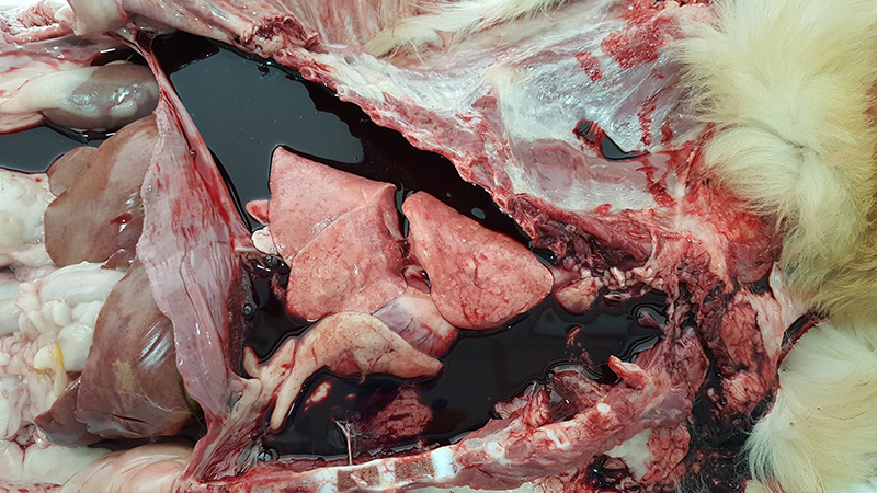
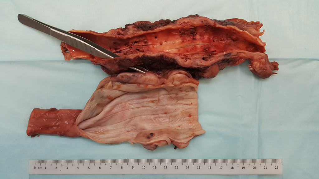
Immediately caudal to the heart base, 14 cm length of the descending aorta was adhered to 11 cm of the immediately adjacent esophagus and formed a fibrous, dark red to white, hemorrhagic mass. Affected segment of descending aorta wall was markedly thickened and had multifocal mural nodules and tunica intima ulceration. There was focal perforation of ventral aortic wall. Tissues and subpleural parenchyma around affected segment of aorta was thickened, hemorrhagic, soft, edematous and friable. The esophageal wall was multifocally thickened by coalescing round to oval nodules. Embedded within the aortic and esophageal nodules were 4-6cm long, 1mm diameter dark red nematodes (Spirocera lupi).
Histopathologic Description:
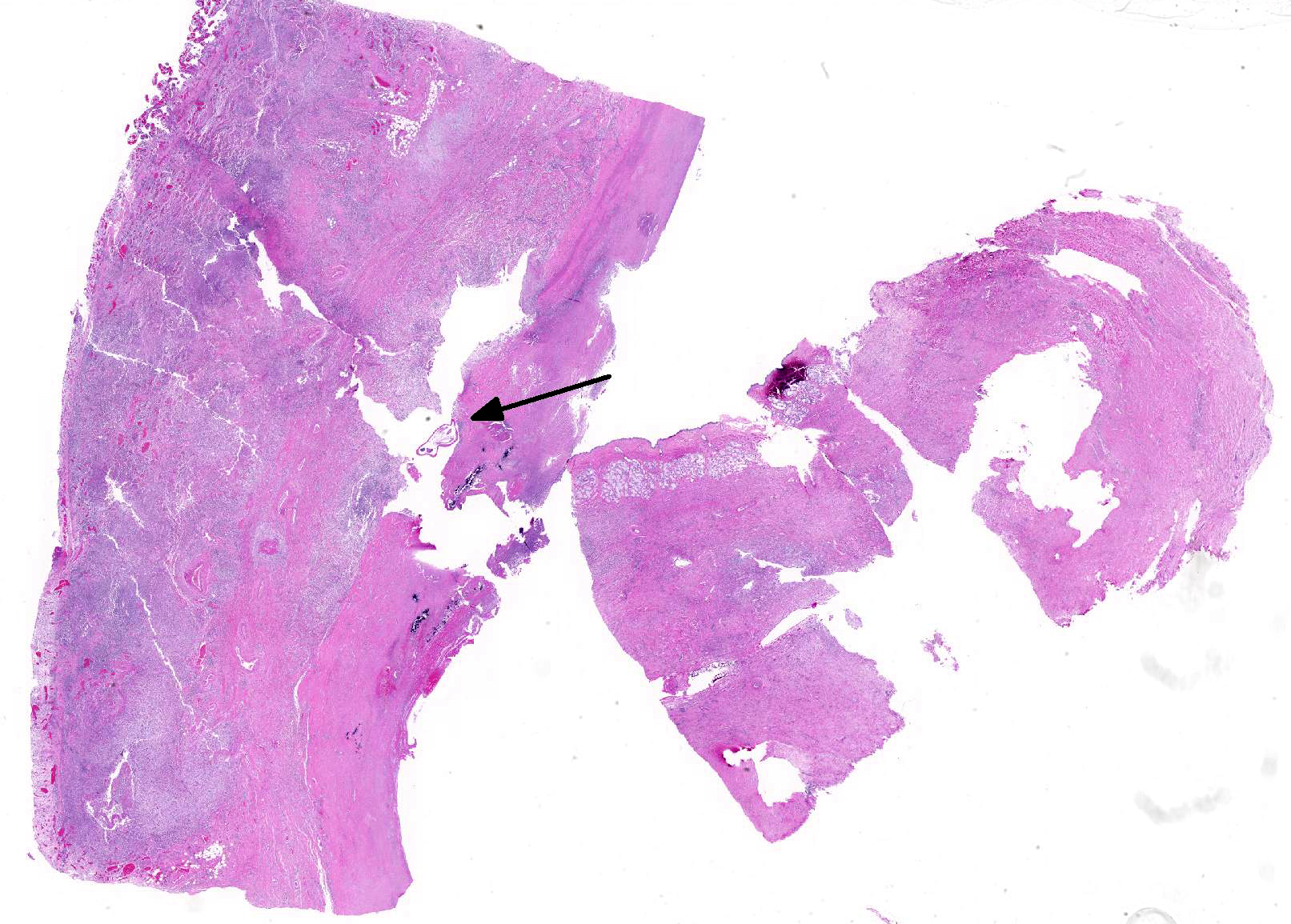
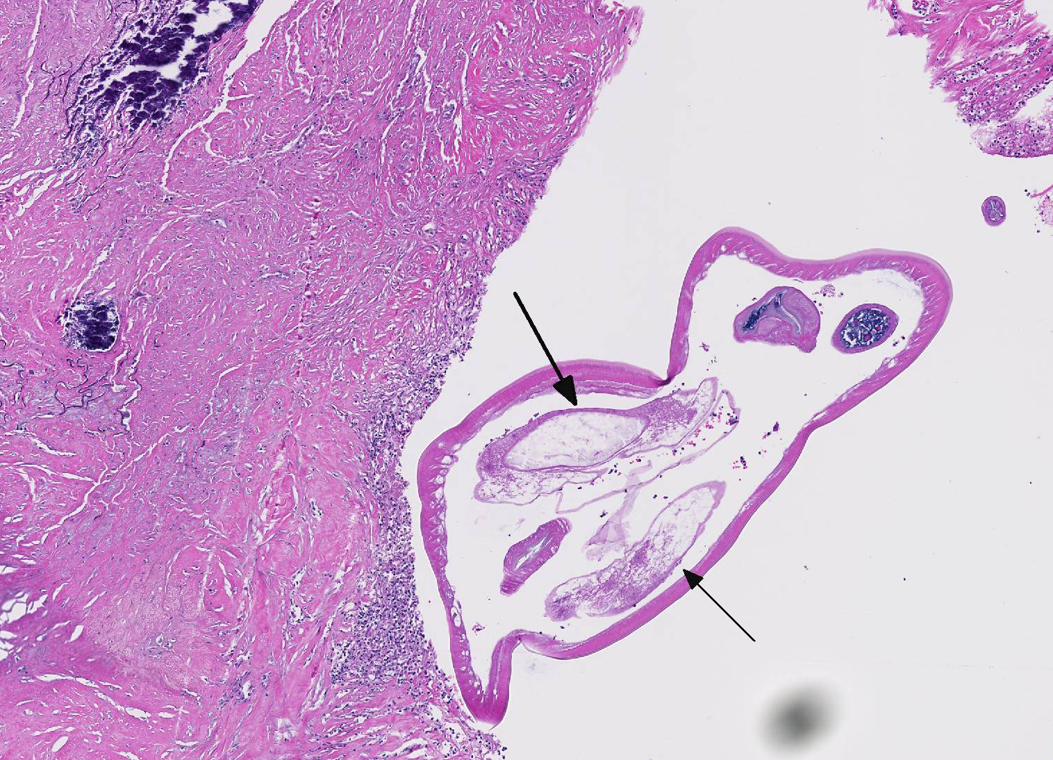
Esophagus : The grossly observed esophagal masses are characterized by expansion of the esophageal wall, mural architecture and replacement by marked necrosis, fibrin exudation and, intense mixed cell infiltrate. The inflammatory cells are intermixed with a large amount of fibrocollagenous stroma, which dissects and replaces tunica muscularis multifocally. Within the necrosis and inflammation, and embedded within esophagus wall are cross sections of 0.2 to 0.3mm diameter nematodes of similar morphology as that described above for the aorta. In a few nodules, among the inflammation and fibrocollagenous stroma are non-encapsulated lobules of atypical and dysplastic mesenchymal cells. Mesenchymal cells are spindle-shaped, arranged in streams, whorls, and bundles supported by loose fibrocollagenous to myxomatous stroma. Mitoses are present at 3 per 10 high power (400x) fields, and anisokaryosis and anisocytosis are both mild. Few scattered nematode eggs are present within the esophageal inflammation, necrosis and atypical mesenchymal cell proliferations.
Morphologic Diagnosis:
Esophagus: 1. Severe, focally extensive, fibrinonecrotizing, eosinophilic, pyo-granulomatous, lymphoplasmacytic eso-phagitis with marked granulating fibrosis, and intralesional Spirurid nematodes (Spirocerca lupi) and ova 2. Multifocal, atypical, dysplastic mesenchymal cell proliferation (fibrosarcoma)
Lab Results:
Condition:
Contributor Comment:
Spirocerca lupi is a spirurid nematode that parasitizes esophageal wall of dogs and some other carnivores and is mostly seen in warm climates where dung beetles act as intermediate hosts. The life cycle of the parasite involves passage of parasite eggs from the definitive host (canine) via feces followed by ingestion by coprophagous (dung-eating) beetle. The larva develops into infective third stage and encysts in the beetles tissues. The beetle is then eaten by a definitive host or by a paratenic host (e.g. lizard, chicken or rodents) which in turn is eaten by the definitive host (dog). The ingested larvae penetrate gastric mucosa and migrate to the aorta via arteries, and embed in the intima aorta and form parasitic granulomas in aortic adventitia at the caudal thoracic area. After a few months, the nematodes migrate to the immediately underlying esophagus, develop to adulthood and form cystic granulomas in distal esophagus submucosa or gastric cardia. Esophageal lesions associated with Spirocerca are characterized by granulomatous inflammation with highly reactive fibroblasts. These foci can develop into fibrosarcoma and osteosarcoma, with local tissue invasion and pulmonary metastasis. The parasites can extend from nodules through fistulas that lead into esophageal lumen, where female worms can oviposit into gastrointestinal tract. Aortic lesions are characterized by necrotizing, hemorrhagic, eosinophilic vasculitis, aneurysm and rare aortic rupture such as in this case. Advanced cases of Spirocerca lupi infection, such as in this case, are associated with guarded to poor prognosis. 2
Aortic rupture due to vasculitis and vascular wall compromise secondary to Spirocerca lupi has been reported to cause a fatal hemothorax in older literature and likely occurred in this case.7 Alternatively, perforation of the esophagus due to damaged esophageal wall from parasitic nodules and/or associated neoplasms can result in leakage of ingesta contents into thoracic cavity and a pyothorax.8
While the majority of the limited land in Singapore consists of built up urban environment, several species of dung beetles still do exist, typically in the remaining forested areas.6,8 These insects, along with wild reptiles and rodents, are the suspected intermediate hosts that infect dogs that come into contact with them in the outdoor environment.
JPC Diagnosis:
2. Aorta: Arteritis, necrogranulomatous and eosinophilic, chronic, diffuse, severe with fibrin thrombi and marked mural fibrosis.
Conference Comment:
Other esophageal parasites discussed by conference participants include Gon-gylonema sp. in ruminants, pigs, horses, primates, and rodents; Gastophilus sp. in horses; and Hypoderma lineatum in ruminants. With the exception of Spirocerca lupi, parasitic diseases of the esophagus are usually of little significance to the host.2,4
Conference participants were unable to reach a consensus regarding the multifocal nodular proliferation of atypical mesenchymal cells within the muscular layers of the esophagus. Some agreed with the contributor, that this likely represents an early neoplastic transformation of the tunica muscularis, while others favored a proliferation of highly activated fibroblasts secondary to chronic marked inflammation. Interestingly, Spirocerca lupi is known for the ability to induce malignant transformation within the wall of its associated esophageal granulomas. Mesenchymal neoplasms fibrosarcoma and osteosarcoma are the most common malignancies associated with the parasite.2,4 Neoplasms that arise from areas of marked granulomatous inflammation are often highly infiltrative with frequent metastasis to the lungs. Additionally, Spirocerca lupi induced thoracic neoplasia has occasionally been associated with the development of hypertrophic osteopathy, a condition that results in the periosteal new bone proliferation in the distal extremities. Other potential sequelae include dysphagia, aortic aneurysm, and aortic rupture leading to acute hemothorax, seen in this case.2,4
Attendees discussed other parasites that induce neoplastic transformation in veterinary species. Residents at the Joint Pathology Center use the acronym SOCCS-T as a mnemonic device to remember which parasites are associated with neoplastic transformation. Spirocerca lupi causes fibrosarcoma and osteosarcoma; Opisthorchis felineus causes cho-langiocarcinoma in cats and humans; Cysticercus fasciolaris can induce hepatic sarcoma in rats; Clonorchis sinensis is associated with cholangiocarcinoma in cats and people; Schistosoma haematobium is a trematode that can induce transitional cell carcinoma of urinary bladder in people; and Trichosomoides crassicauda can cause papillomas of the urothelium in rats.2-4
References:
- Blume GR, Reis JL, Gardiner CH, et al. Spirocerca lupi granulomatous pneumonia in two free-ranging maned wolves (Chrysocyon brachyurus) from central Brazil. J Vet Diagn Invest. 2014; 26(6):815-817.
- Uzal FA, Plattner BA, Hotetter JM. Alimentary system. In: Maxie MG, ed. Jubb, Kennedy and Palmers Pathology of Domestic Animals. Vol 2. 6th ed. Philadelphia, PA: Elsevier Saunders; 2016:34-35.
- Gardiner CH, Poynton SL. Spirurids. In: Gardiner CH, Poynton SL, eds. An Atlas of Metazoan Parasites in Animal Tissues. Washington, DC: Armed Forces Institute of Pathology; 1999:30-34.
- Gelberg HB. Alimentary system and the peritoneum, omentum, mesentery, and peritoneal cavity. In: In: Zachary JF, ed. Pathologic Basis of Veterinary Disease. 6th ed. St. Louis, MO: Elsevier Mosby; 2017:356-357.
- Klainbart S et al. Spirocercosis-associated pyothorax in dogs. Vet J. 2007; 173(1):209-214.
- Lee JS, Lee IQW, Lim SL, et al. Changes in dung beetle communities along a gradient of tropical forest disturbance in South-East Asia. J Tropic Ecol. 2009; 25(6):677-680.
- Rinas MA, Nesnek R, Kinsella JM, DeMatteo KE. Fatal aortic aneurysm and rupture in a neotropical bush dog (Speothos venaticus) caused by Spirocerca lupi. Vet Parasitol. 2009; 164(2-4):347-9.
- Xin RO, Siew CC, Potts MD. Recent records of the dung beetle Catharsius Molossus (Coleoptera: Scarabaeidae) in Singapore. Nature in Singapore. 2013; 19(6): 16.