Joint Pathology Center
Veterinary Pathology Services
Wednesday Slide Conference
2017-2018
Conference 3
September 6th, 2017
CASE II: 11-30435 (JPC 4020992).
Signalment: 2-week-old male and female intact, terrier mixed breed dogs (Canis lupus familiaris).
History: An adult terrier mixed breed dog was vaccinated with canine distemper virus during pregnancy, and 7 out of 8 puppies died after birth. Upon initial presentation, 6 puppies had normal physical exams and normal temperatures, while the 7th puppy was vocalizing, dyspneic, mildly cyanotic, and had a mildly elevated lymphocyte count. This puppy was euthanized a short time later. The rest of the puppies had intermittent twitching, one puppy was hypoglycemic, and one puppy had moderate amounts of clear nasal discharge. The rest of the puppies died within the next few days.
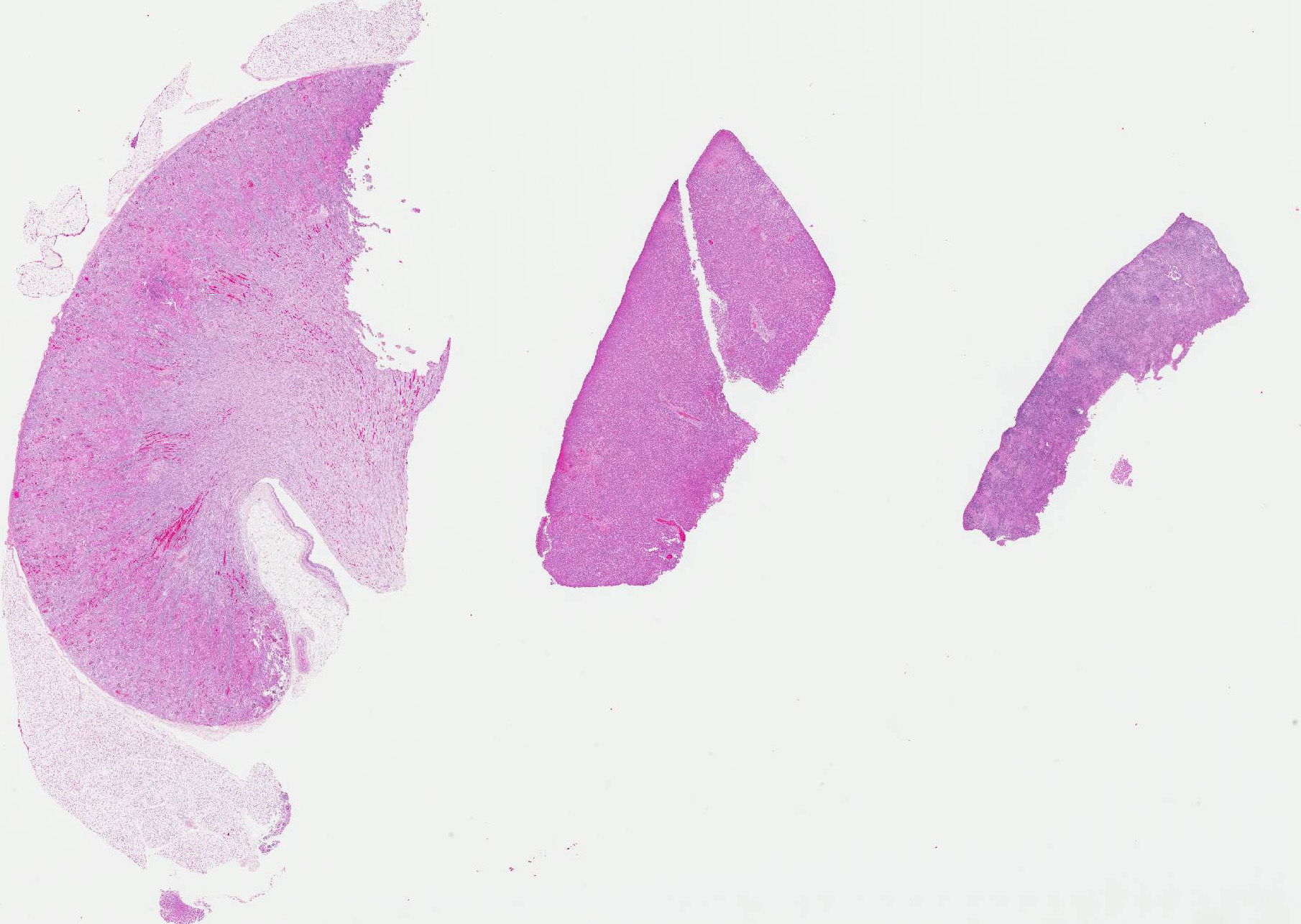
Gross Pathology: The kidney, liver, spleen, and to a lesser extent within the lungs and small intestines were multifocal pinpoint to 1 mm, dark red foci (petechia). The thoracic cavity contained approximately 5-10 ml of yellow to red-tinged, translucent fluid. The lungs were diffusely red and rubbery.
Laboratory results: FA was positive for canine herpesvirus.
FA was negative for canine distemper virus.
Bacteriology showed no growth in the lung and kidney.
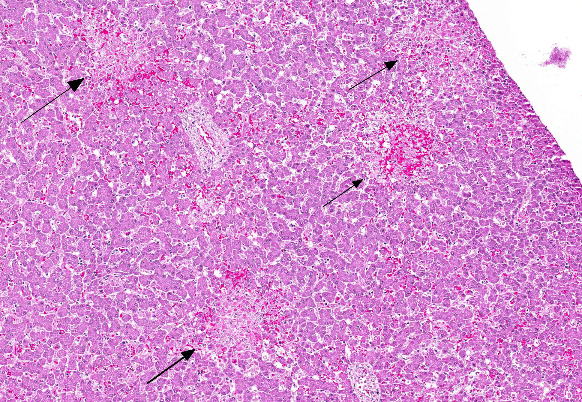
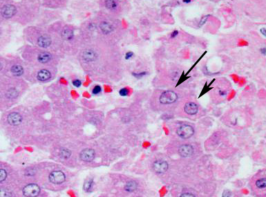
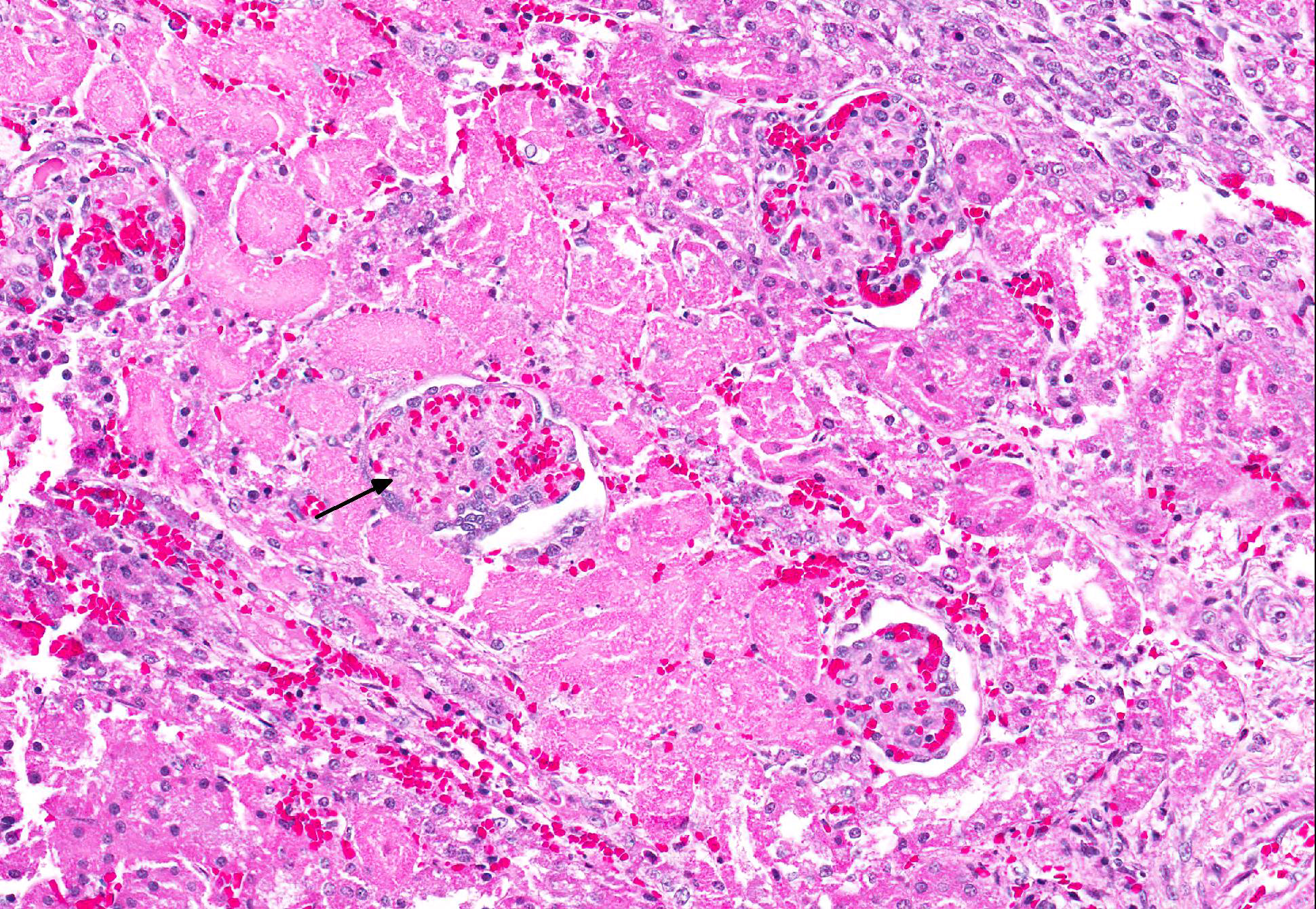
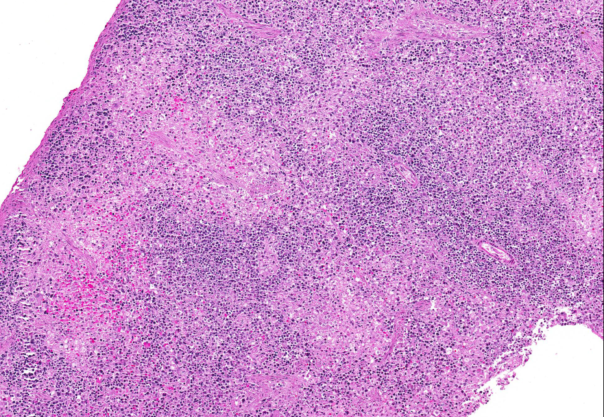
Microscopic Description: Multifocally within sections of kidney, liver, and spleen, the parenchyma has lost differential nuclear staining and contains necrotic cellular debris and hemorrhage. The surrounding cells often have karyorrhectic nuclei with hypereosinophilic cytoplasm. Few nuclei contain a single, round, eosinophilic intranuclear inclusion bodies with marginated chromatin.
Additionally, tissue from the lungs and jejunum (slides not submitted) have similar multifocal regions of necrosis with few single, round, eosinophilic intranuclear inclusion bodies with marginated chromatin. The alveolar septa are also diffusely expanded by macrophages, lymphocytes, and neutrophils with type II pneumocyte hyperplasia. The grey and white matter of the cerebrum, cerebellum, and brainstem (slides not submitted) are multifocally disrupted by glial cells, macrophages, few neutrophils, and degenerating neurons. The meninges are diffusely thickened with neutrophils, lymphocytes, and macrophages.
lmmunohistochemistry: The small intestine was negative for canine parvovirus, and the brain was negative for canine distemper virus.
Contributors Morphologic Diagnosis: Kidney, liver, and spleen: Nephritis, hepatitis, and splenitis, necrotizing, multifocal, marked, with few eosinophilic intranuclear inclusion bodies.
Etiology: Canine herpesvirus-1 (CaHV-1)
Contributors Comment: Canine herpesvirus type 1 is a well-known entity first recognized in the 1960s and is a double stranded DNA virus in the alphaherpesvirus subfamily. The virus is deactivated in temperatures greater than 40°C and flourishes in temperatures between 34°C and 36°C; thus, by raising the body temperature, chances of survival are increased.2, 5, 7
The age of the dog can determine the clinical presentation of the herpesvirus infection. Dogs less than 3 weeks old generally have fatal systemic disease due to acute neonatal viremia. Dogs older than 3 weeks can have ocular and mucosal disease (either respiratory or vaginal). Naïve pregnant bitches are susceptible to systemic infection that can cause abortions or acute neonatal viremia. Latent infection is a concern for any age group if they survive the original infection and can occur following steroid administration or stress, such as pregnancy.1, 2, 3
Typically, dogs less than 3 weeks of age acquire the disease in a variety of methods: in utero, passage through the birth canal, direct contact with oronasal secretions of the bitch, direct contact with other dogs that are shedding, and humans acting as fomites. Older dogs with ocular and/or mucosal disease are generally infected via aerosolization. Viral replication can occur in the nasopharynx, tonsils, and/or retropharyngeal and tracheobronchial lymph nodes. The virus then spreads throughout the bloodstream via macrophages to infect the liver, kidneys, lymph nodes, lungs, and centralnervous system.1, 2, 3, 7 The incubation period is approximately 6 to 10 days and litter mortality can reach 100% over the course of a week.2 Common clinical signs include vocalization, anorexia, hypothermia, abdominal pain, and/or incoordination.2, 4
Characteristic gross and histologic lesions in affected dogs less than 3 weeks of age include systemic hemorrhage and necrosis in the kidney, liver, spleen, and intestine with little associated inflammation, enlarged lymph nodes, and non-suppurative meningoencephalitis, all of which were present within this case.2, 4, 5, 6, 7
Eosinophilic intranuclear inclusion bodies were also present. Classic lesions in addition to a positive fluorescent antibody test verified canine herpesvirus. Fluorescent antibody testing and immunohistochemistry ruled out canine distemper virus and canine parvovirus as possible contributors.
JPC Diagnoses: 1. Liver: Hepatitis, necrotizing, random, multifocal to coalescing with few eosinophilic intranuclear inclusion bodies, terrier mix, canine.
2. Kidney, tubules and glomeruli: Nephritis, necrotizing, multifocal, mild.
3. Spleen: Splenitis, necrotizing, diffuse, severe.
Conference Comment: This case serves as a classic example of canine herpesvirus-1 (CaHV-1) in a puppy, causing random necrotizing hepatitis, nephritis, and splenitis. Participants described multifocal areas of coagulative necrosis in the liver and kidney, as well as diffuse necrosis in the spleen, with few eosinophilic intranuclear inclusion bodies, mild vasculitis, and fibrin thrombi (especially in the liver and glomeruli). Participants also noted that inclusions were most prominent in the liver adjacent to areas of necrosis, but were difficult to see in the kidney and spleen due to the extensive necrosis in those tissues.
In addition, participants reviewed select alphaherpesviruses from other species (cats, farm animals, and horses), which are summarized in the following chart1, 3, 7, 8, 11:
|
Alphaherpesvirus |
Common name |
Primary lesions |
|
Bovine herpesvirus 1 |
Infectious bovine rhinotracheitis |
Fibrinonecrotic membrane along trachea/larynx, bronchointerstitial pneumonia; Systemic form: Focal areas of necrosis within the alimentary tract |
|
Infectious pustular vulvovaginitis |
Necrotizing vulvovaginitis with intranuclear inclusion bodies in epithelial cells |
|
|
Bovine herpesvirus 2 |
Pseudo-lumpy skin disease |
Eruption of superficial cutaneous nodules with a depressed center; no scar formation or deep necrotic sequestra (differentiates from true lumpy skin disease) |
|
Bovine mammillitis |
Trauma initiates infection; swollen teats with cutaneous plaques; epithelial syncytia with Cowdry type A intranuclear viral inclusions |
|
|
Porcine herpesvirus 1 |
Pseudorabies/Aujeszkys Disease |
Abortion; non-suppurative meningoencephalitis; rhinitis, tonsillitis, pneumonia; coagulative necrosis of placenta, liver, spleen, adrenal gland |
|
Equine herpesvirus 3 |
Equine coital exanthema |
Papules, vesicles, pustules on genitalia or muzzles; ballooning degeneration of keratinocytes with intranuclear viral inclusions |
|
Equine herpesvirus 5 |
Multinodular pulmonary fibrosis |
Nodules with thick interstitial collagen with irregular alveoli lined by cuboidal cells; intranuclear viral inclusions in alveolar macrophages |
|
Gallid herpesvirus 1 |
Avian infectious laryngotracheitis |
Mucohemorrhagic fibrinonecrotic exudates (+/- tracheal casts) within nasal turbinates, sinuses, conjunctiva, larynx, and trachea; epithelial ulceration with formation of syncytial cells containing intranuclear viral inclusions |
|
Gallid herpesvirus 2 |
Mareks disease |
Herpesviral induced T-cell lymphoma; four different gross lesion patterns: (1) thickening and yellowing of peripheral nerves (2) discoloration of the iris (3) enlargement of feather follicles and (4) visceral tumors |
|
Anatid herpesvirus 1 |
Duck Plague |
Multifocal hemorrhages in visceral organs; severe enteritis with lymphoid tissue necrosis; foci of necrosis in liver |
|
Feline herpesvirus 1 |
Feline viral rhinotracheitis |
Focal ulcerative (and often eosinophilic) lesions on face or nasal planum (usually without clinical respiratory signs); ulcerative and necrotizing with large glassy intranuclear viral inclusions in epithelial cells |
Contributing Institution:
University of Illinois
College of Veterinary Medicine
Veterinary Diagnostic Laboratory
Urbana, IL 61802
References:
- Cantile C, Youssef S. Nervous system. In: Maxie MG, ed. Jubb, Kennedy, and Palmers Pathology of Domestic Animals. Vol 1. 6th ed. St. Louis, MO: Elsevier; 2016:382-383.
- Carmichael LE, Greene CE. Canine herpesvirus infection. In: Greene CE, ed. Infectious Diseases of the Dog and Cat. 3rd ed. Elsevier; 2006: 47-53.
- Caswell JL, Williams KJ. Respiratory system. In: Maxie MG, ed. Jubb, Kennedy, and Palmers Pathology of Domestic Animals. Vol 2. 6th ed. St. Louis, MO: Elsevier; 2016:537-538, 568, 577.
- Decaro N, Martella V, Buonavoglia C. Canine Adenoviruses and Herpesvirus. Vet Clin North Am Small Anim Pract. 2008; 38 (4): 799-814.
- Evermann JF, Ledbetter EC, Maes RK. Canine reproductive, respiratory, and ocular diseases due to canine Herpesvirus. Vet Clin North Am Small Anim Pract. 2011; 41 (6): 1097-1120.
- Kojima A, Fujinami F, Takeshita M, Minato Y, Yamamura T, Imaizumi K, Okaniwa, A. Outbreak of neonatal canine Herpesvirus infection in a specific pathogen free beagle colony. Jpn J Vet Sci. 1990; 52 (1 ): 145-154.
- Mauldin EA, Peters-Kennedy J. Integumentary system. In: Maxie MG, ed. Jubb, Kennedy, and Palmers Pathology of Domestic Animals. Vol 1. 6th ed. St. Louis, MO: Elsevier; 2016:625-627.
- Ojkic D, Brash ML, Jackwood MW, Shivaprasad HL. Viral diseases. In: Boulianne M ed. Avian Disease Manual. 7th ed. Jacksonville, FL: American Association of Avian Pathologists, Inc.; 2013:30-31, 42-43, 59-60.
- Percy DH, Carmichael LE, Albert DM, King JM, Jonas AM. Lesions in puppies surviving infection with canine Herpesvirus. Vet Path. 1971; 8: 37-53.
- Percy DH, Olander HJ, Carmichael LE. Encephalitis in the newborn pup due to a canine Herpesvirus. Vet Path. 1968; 5: 135-145.
- Schlafer DH, Foster RA. Female genital system. In: Maxie MG, ed. Jubb, Kennedy, and Palmers Pathology of Domestic Animals. Vol 3. 6th ed. St. Louis, MO: Elsevier; 2016:431-432, 443-445.
- Zachary JF, McGavin MD. Pathologic Basis of Veterinary Disease. 5th ed. Elsevier; 2012:218, 654, 1117.