Joint Pathology Center
Veterinary Pathology Services
Wednesday Slide Conference
2011-2012
Conference 25
16 May 2012
CASE III: 10-2049 (JPC 4004362).
Signalment: 10-month-old male castrated Scottish terrier dog (Canis familiaris).
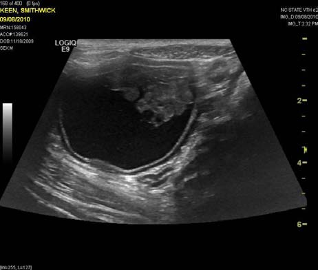
History: The dog was diagnosed with a urinary tract infection 4 months prior to presentation, and was treated with antibiotics. The infection resolved but there was ongoing, waxing and waning hematuria. At the time of presentation, there had been a recent onset of polyuria and increased urgency to urinate. Contrast radiographs at the time of presentation revealed a filling defect in the urinary bladder. Ultrasound imaging showed a broad-based, heteroechoic mass arising near the trigone region of the bladder. A portion of the urinary bladder was surgically resected and submitted for histologic evaluation.
Gross Pathology: A segment of bladder wall approximately 2 x 3 cm was submitted for biopsy with an attached 2 x 4 x 3 cm, polypoid, soft, red-brown mass that protruded into the lumen of the bladder. Approximately 2 cm of ureter were also submitted.
Laboratory Results: CBC:
WBC 18,100/ul
Neutrophils 14,118/ul
Lymphocytes 543/ul
Monocytes 1,629/ul
Serum Chemistry:
Albumin 2.6 g/dl (N: 3.0-3.9)
Urinalysis:
Albumin 500 ug/dl (3+)
Blood 250/ul
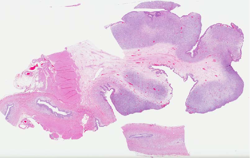
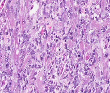
Contributorâs Histopathologic Description: Multiple histologic sections were examined with similar findings. Arising from the mucosal epithelium is a multilobular mass supported by a fibrovascular core. Lobules of the mass are composed of a background of neoplastic spindle cells with a myxomatous stroma admixed with frequent multi-nucleated, large, elongate or rounded cells with abundant, fibrillar, eosinophilic cytoplasm. The elongate cells occasionally have prominent cross-striations and are consistent with myogenic âstrap cells.â The multiple nuclei of the elongate and large round cells have peripheralized chromatin with 1-2 prominent nucleoli. The spindle cells in the background have indistinct cell borders with scant to moderate pale eosinophilic cytoplasm around a central oval nucleus. The nuclei have coarsely clumped to vesicular chromatin and often have 1-2 prominent nucleoli. There is marked anisocytosis and anisokaryosis. Mitotic figures are observed with 14 per 10 high power fields. Each lobule of the mass is supported by a fibrovascular core and is lined by transitional epithelium. The ureter has hyperplastic epithelium, and there appears to be a portion of a neoplastic lobule that is within the ureteral lumen, but not attached to the wall of the ureter.
Contributorâs Morphologic Diagnosis: Urinary bladder: Botryoid rhabdomyosarcoma.
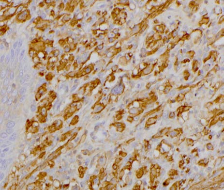
Contributorâs Comment: Botryoid rhabdomyosarcomas are rare tumors in dogs that most frequently occur in the urinary bladder of young, large breed dogs. There are four categories of rhabdomyosarcomas that are recognized in people, including embryonal, botryoid, alveolar, and pleomorphic. Embryonal rhabdomyosarcomas are characterized by the presence of primitive myogenic cells that occur in two forms, either with large, well-differentiated rhabdomyoblasts on a background of smaller round cells, or a myotubular arrangement with spindle cells forming a myxoid arrangement and often with multinucleate cells and strap-like cells that may or may not have cross striations. Botryoid rhabdomyosarcomas are generally regarded as a variant of embryonal rhabdomyosarcoma with a histologic appearance consistent with the myotubular form, and a gross appearance of a polypoid, grape-like pattern, that often projects into the lumen. The presence of myogenic cells is diagnostic, and myogenic origin can be confirmed with a number of immunohistochemical markers including desmin, muscle-specific actin, and myoglobin. In addition, myogenin and MyoD can be used to identify specific stages of myogenesis.7 PTAH stains can also be used to try to recognize cross-striations if they are present. In this case, the prominent multinucleated and strap-like cells are diagnostic. In addition, there is strong immunohistochemical staining for muscle specific actin.
Saint Bernards are often listed as being overrepresented in the occurrence of botryoid rhabdomyosarcomas based on a 1973 paper with a case series in which 4 of 7 dogs were Saint Bernards.5 However, in a review of published cases of canine botryoid rhabdomyosarcomas, no additional cases in Saint Bernards have been described, and there have been at least three cases reported in golden retrievers.1,7 Other breeds affected include Labrador retriever, Maltese, Newfoundland, Rottweiler, Basset hound, miniature poodle, and a great Dane.1,5,6,8,9,10 In addition to occurring in the urinary bladder, a few case reports have described botryoid rhabdomyosarcomas in the genital tract as well,1,9 and there is also a case report of an embryonal rhabdomyosarcoma in the oropharynx and temporal muscles of a young Bassett hound.6
Botryoid rhabdomyosarcomas typically have a poor prognosis. The metastatic rate is not well-reported as several cases are euthanized close to the time of initial diagnosis or there is a lack of follow-up in many cases. Out of 14 cases of genital or urinary tract botryoid rhabdomyosarcomas described in case reports, at least five of them had widespread metastases to multiple organs including liver, lungs, lymph nodes, heart, subcutaneous tissue, muscle, gingiva, adrenal gland, spleen, and kidney.1,5,6,10
JPC Diagnosis: Urinary bladder: Botryoid rhabdomyosarcoma.
Conference Comment: The contributor provided an excellent discussion of botryoid rhabdomyosarcoma and the associated pathology. Conference participants discussed the following common clinical sequella to this neoplasm. Urinary outflow obstruction is a common finding with these neoplasms, resulting in post-renal azotemia, hematuria, dysuria, and stranguria. The proteinuria and large amount of albumin in the urine can be attributed to the hematuria. Hypertrophic osteopathy is reported to occur concurrently with botryoid rhabdomyosarcoma, thought to be due to vascular or neurogenic stimuli which affect changes in the peripheral vascular supply, as seen with pulmonary masses. Poorly oxygenated blood passes through arteriovenous shunts, producing local passive congestion and poor tissue oxygenation and stimulating proliferation connective tissue, including the periosteum and the synovial membrane. The affected animals are usually young and present bilaterally symmetrical, nonedematous soft tissue swellings affecting primarily the distal portions of all four limbs. The initial soft tissue swellings are soon accompanied by a diffuse periosteal new-bone formation, which may ultimately affect all the bones of the limbs, and the bone lesions resolve after tumor removal.2,3,4
Contributor: N.C. State University
College of Veterinary Medicine
Population Health and Pathobiology-Histology Lab
1060 William Moore Dr.
Raleigh, NC 27607
http://www.cvm.ncsu.edu/dphp/path/anatomicpath.html
References:
1. Bae IH, Kim Y, Pakhrin B, et al. Genitourinary rhabdomyosarcoma with systemic metastasis in a young dog. Vet Pathol 2007;44:518-520.
2. Brodey R. Hypertrophic osteoarthropathy in the dog: A clinicopathologic study of 60 cases. J Am Vet Med Assoc. 1971;159:1242.
3. Cooper BJ, Valentine BA. Tumors of muscle. In: Meuten DJ, ed. Tumors of Domestic Animals. 4th ed. Ames, IA: Iowa State Press; 2002:354.
4. Halliwell WH, Ackeman N. Botryoid rhabdomyosarcoma of the urinary bladder and hyperosteoarthropathy in a young dog. J Am Vet Med Assoc. 1974;165:911.
5. Kelly DF. Rhabdomyosarcoma of the urinary bladder in dogs. Vet Pathol. 1973;10:375-384..
6. Kim DY, Hodgin EC, Cho DY, et al. Juvenile rhabdomyosarcomas in two dogs. Vet Pathol. 1996;33:447-450.
7. Kobayashi M, Sakai H, Hirata A, et al. Expression of myogenic regulating factors, Myogenin and MyoD, in two canine botryoid rhabdomyosarcomas. Vet Pathol. 2004;41:275-277.
8. Kuwamura M, Yoshida H, Yamate J, et al. Urinary bladder rhabdomyosarcoma (sarcoma botryoides) in a young Newfoundland dog. J Vet Med Sci. 1998;60:619-621.
9. Suzuki K, Nakatani K, Shibuya H, et al. Vaginal rhabdomyosarcoma in a dog. Vet Pathol. 2006;43:186-188.
10. Takiguchi M, Watanabe T, Okada H, et al. Rhabdomyosarcoma (botryoid sarcoma) of the urinary bladder in a Maltese. J Small Anim Pract. 2002;43:269-271.