Signalment:
25-year-old Quarter Horse gelding,
Equus caballus, equine.One of three horses on a ranch in Southern California was found within a week circling and walking aimlessly. According to the submitting veterinarian this horse had elevated liver enzymes (specific enzymes and their values were not provided) and it was treated for a week for hepatic encephalopathy (no further details provided), after which it was euthanized due to lack of response to treatment.
Gross Description:
The carcass was in poor nutritional condition, with no fat reserves, sunken eyes and mild generalized muscle atrophy. The cardiac and bone marrow (femur) fat showed severe and diffuse serous atrophy. There was a moderate amount (occupying approximately 1/5 of the lumen) of coarse sand, mixed with digesta in all four sections of the large colon. The cecum contained a large amount of reddish, translucent fluid, although no significant gross abnormalities were observed on the mucosa of this organ. The liver was reduced in size (approximately 1/3 to 1/2 of its normal size) and thickness; the consistency was, however, unremarkable. No other significant gross abnormalities were observed in the rest of the carcass.Â
Histopathologic Description:
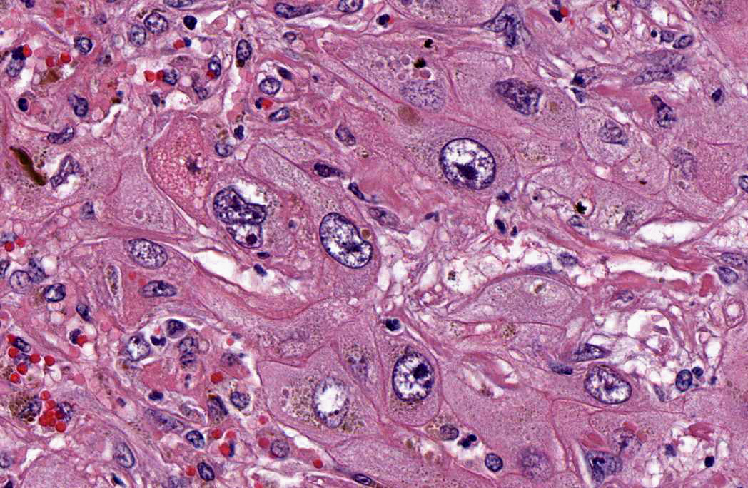
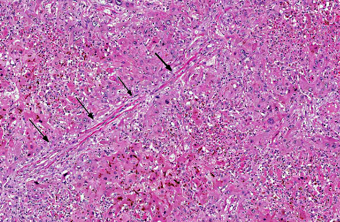
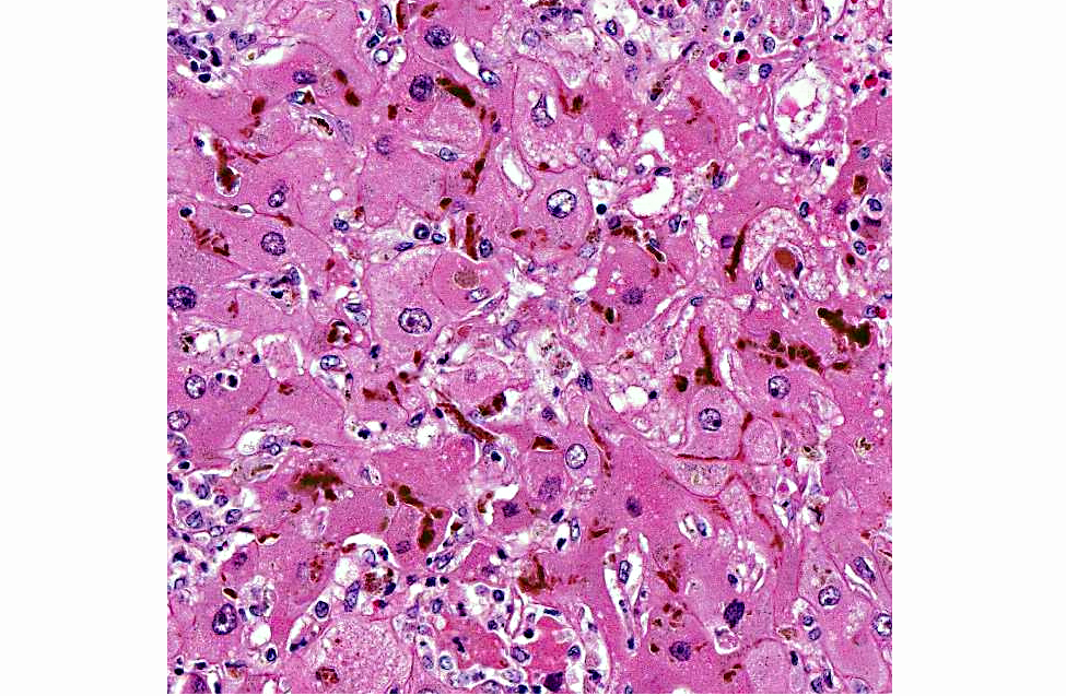
Diffusely, there is severe megalocytosis consisting of enlargement of the nuclei and cytoplasm of a high percentage of hepatocytes. The volume of affected cells is increased as much as to 5 to 10 times. The nuclear membrane is basophilic and sharply delimited. Most hepatocytes have one nucleolus, although up to four nucleoli are present in some cells. The chromatin is fragmented. The peripheral part of the cytoplasm is usually pale and presents multiple coalescing vacuoles. Acidophilic, more or less spherical bodies (cytosegresomes), are occasionally seen in the cytoplasm of some hepatocytes. Because the enlarged hepatocytes are closely apposed, the sinusoids are rarely evident and the general architecture of the organ is distorted. Small to medium size pools of bile (bile stasis) are present in canaliculi and in the cytoplasm of hepatocytes. Multifocally, individual cell necrosis is observed. Diffusely, the liver presents moderate fibrosis, which is mostly restricted to the portal areas, although some fibrosis can be seen attempting to dissect lobules and separate individual cells. Multifocally, there is bile duct proliferation with hypertrophic epithelium. Multifocally there is hemorrhage, and infiltration of neutrophils, lymphocytes, plasma cells and macrophages. Blood vessels in portal areas show plump, hypertrophic epithelium. These changes tend to affect all the cells of all the lobules with no special predilection for any part of the lobule.Â
In the brain (not submitted) there are single or small groups of astrocytes with clear, vesicular nuclei (Alzheimer type II cells), with scant cytoplasm, mostly restricted to basal nuclei, cerebellar peduncles and hippocampus.Â
Morphologic Diagnosis:
Hepatopathy, with megalocytosis, portal fibrosis, bile duct proliferation and bile stasis, diffuse, chronic.
Lab Results:
Salmonella culture of liver: negative; Aerobic culture of liver: no growth in 48 hours; Anaerobic culture of colon content: no growth in 48 hours;
C. difficile culture (colon content): negative; Fecal float: no parasite eggs seen;
C. perfringens toxins (alpha, beta, epsilon) ELISA in small intestine and colon content: negative;
C. difficile toxins A/B ELISA in small intestine and colon content: negative; Brain cholinesterase activity: within normal range; Heavy metal screen in liver (including selenium): within normal range. Examination of a bale of alfalfa hay from a batch that this horse and his companions had been eating for the past several months identified the following plants: common groundsel, cheeseweed, wild lettuce, shepherds purse and green foxtail.Â
Condition:
Senecio vulgaris
Contributor Comment:
The changes described in the liver are classic of pyrrolizidine alkaloids (PA) intoxication, which is consistent with the presence of common groundsel (
Senecio vulgarism), a plant known to contain PAs, in the hay that this animal was eating.(4) PAs are hepatotoxic, causing irreversible liver damage. Horses and cattle are the major livestock species affected by PAs. Clinical signs of chronic PA poisoning may often not appear for 2-8 months after the first ingestion of PA-containing plants. Affected animals lose condition, and develop liver failure. Neurological signs are commonly seen in horses, and the condition is called "walking disease".(2) After the onset of clinical signs, the prognosis is poor. The most characteristic effect of PA on the liver is the induction of megalocytosis, due to an antimitotic effect. Megalocytosis is not due to inhibition of DNA synthesis, as continued nucleoprotein synthesis, combined with mitotic inhibition, accounts for the increase in size of the nuclei. Although megalocytosis is a hallmark of PA intoxication, this change is not specific for this intoxication, as it can be seen in intoxication by aflatoxins and nitrosamines as well. In this case, however, the hepatic lesions (including megalocytosis, fibrosis and bile duct proliferation) coupled with the detection of Senecio vulgaris in the hay that this horse was consuming, is compelling evidence that this horse suffered from PA intoxication.(4) Although PAs can be tested for gastrointestinal contents, the fact that the lesions were so chronic makes testing of doubtful value as it was likely that any PA would have been metabolized long before this animal died. This horse also had neurological clinical signs and presence of Alzheimer type II cells in the brain. These clinical signs and lesions are characteristic of hepatic encephalopathy, a frequent complication of liver failure due to PA intoxication in horses and other animal species. While hepatic encephalopathy in several animal species is characterized by the presence of Alzheimer type II cells and spongiform change of white matter, in the horse, changes are limited to the development of type II cells, which was the case with this animal.(2)
Several toxic plants were identified in the bale of hay belonging to the batch that this animal was eating. Amongst those, the most significant is
Senecio vulgaris (common groundsel), which was most likely responsible for the hepatic lesions and related clinical signs observed in this animal.Â
Senecio vulgaris is a common weed in hayfields in California and is also widely distributed along the East Coast and Canada. Identification of PA-containing weeds in alfalfa and detection of PAs in forage are important to establish an accurate diagnosis.(1) Other plants found in the hay bale are also considered to be toxic, although they probably did not play a role in the disease of this horse. Amongst these, cheeseweed (
Malva parviflora) is suspected to cause tremors (i.e. shivers or staggers), which may be intensified by exercise, followed by prostration and death. The causative agent is thought to be malvalic and sterculic acid. Wild Lettuce (
Lactuca virosa) is reported to cause opium-like symptoms. It possesses mild sedative and hypnotic properties. Shepherds purse (
Capsella bursa-pastoris) belongs to the mustard family and is reported to cause congenital hypothyroid dysmaturity syndrome in foals. Green foxtail (
Setaria spp.) is among many species of grass with long, stiff awns or bristles that can cause physical injury to animals. The bristles can puncture sensitive tissue in the mouth and around the nose or eyes. The minutely barbed awns or bristles can work further into a wound by the movement of the animal.(1)
JPC Diagnosis:
Liver: Hepatocellular degeneration, necrosis and loss, diffuse, severe, with megalocytosis, bridging portal fibrosis and cholestasis.
Conference Comment:
The contributor provides a very good description of clinical and histologic manifestations of pyrrolizidine alkaloid intoxication. There are many PA-containing plants, including species in the
Senecio, Crotalaria, Cynoglossum, Echium, Heliotropium, Amsinckia, Symhytum, Borago, and
Trichodesma genera.(3) Pyrrolizidine alkaloids are composed of free base and N-oxides. Although not directly toxic, once these alkaloids are bioactivated by mixed function oxidases, primarily in centrilobular regions of the liver, the resulting pyrroles are potent electrophiles that bind to and cross-link DNA, proteins, amino acids and glutathione. This results in both cytotoxic and antimitotic effects on hepatocytes. Plant species vary in the amount and variety of PAs they contain; hence the resulting pyrroles vary as well, with some being significantly more hepatotoxic than others. For example, the metabolites of seneciphylline and retrorsine are primarily hepatotoxic, whereas less reactive PAs such as trichodesmine and monocrotaline result in fewer hepatic changes and more extensive extrahepatic lesions. Animal species vary significantly in their susceptibility to toxic effects, likely due to variation in metabolism. For example, the toxic dose of some PA containing plants is 20 times higher for sheep than that for cattle. Relative susceptibility to PA poisoning in domestic animals are: pigs=1 (most sensitive); chickens =5; cattle and horses =14; rats = 50; mice =150; sheep and goats=200 (least sensitive). Additionally, the age, nutrition status and gender of the animal are also important factors in determining the severity of disease. Young animals and animals with suboptimal nutrition are at higher risk, and studies have found male rats are more susceptible than females.(3)
Early cellular changes in PA intoxication are dose-dependent hepatocyte swelling which progresses to degeneration and necrosis. Quickly-ingested high doses result in acute intoxication, characterized by panlobular hepatocellular necrosis, hemorrhage and minimal inflammation. Serum clinicopathologic abnormalities in acute toxicity include elevations in aspartate aminotransferase (AST), sorbitol dehydrogenase (SDH), alkaline phosphatase (ALK), and gamma-glutamyl transpeptidase (GGT) as well as increased bilirubin and bile acids. When lower doses are ingested over a longer period of time, chronic poisoning results. Early chronic lesions include piecemeal hepatic necrosis, minimal peribiliary fibrosis and mild bile duct regeneration. With time, megalocytosis and fibrosis occurs, as in this case, and animals eventually develop clinical liver failure and sequalae such as photosensitization, icterus, and increased susceptibility to hepatic lipidosis and ketosis. In chronic cases, animals may develop transient elevations of serum enzymes, bilirubin and bile acids; however, these may remain within normal limits for months after the PA ingestion.(3)
References:
1. Burrows GE, Tyrl RJ.Â
Toxic plants of North America. Ames, Iowa: Iowa State University Press; 2001.
2. Kelly WR. The liver and biliary system. In: Jubb KVF, Kennedy PC, Palmer N.Â
Pathology of Domestic Animals. Vol. 2. London, UK: Academic Press; 1993:319-406.
3. Stegelmeier BL. Pyrrolizidine Alkaloidcontaining toxic plants (
Senecio, Crotalaria, Cynoglossum, Amsinckia, Heliotropium, and
Echium spp.).Â
Vet Clin Food Anim. 2011;27:419428.
4. Summers BA, Cummings JF, de Lahunta A. Degenerative diseases of the central nervous system. In:
Veterinary Neuropathology. St. Louis, Missouri: Mosby; 1995:208-350.