Signalment:
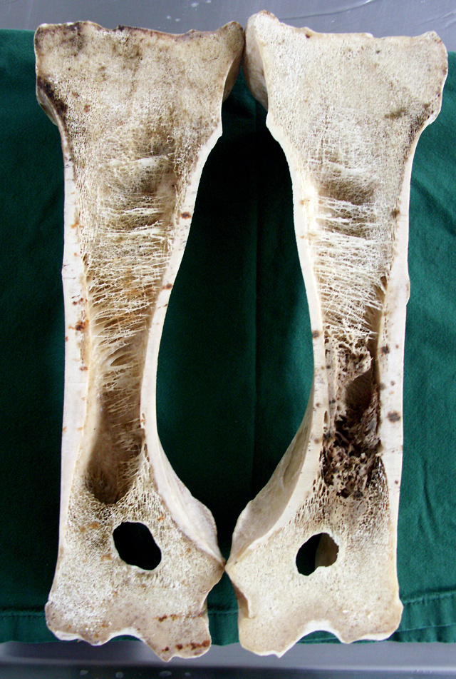
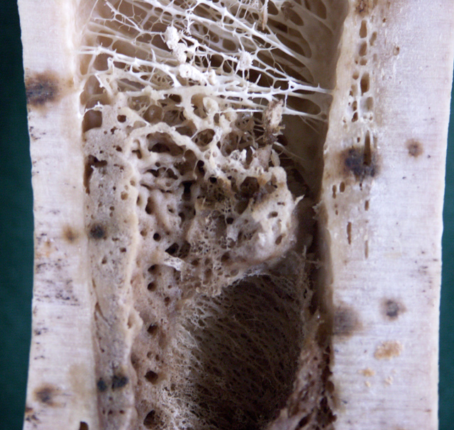
Gross Description:
Histopathologic Description:
Morphologic Diagnosis:
Condition:
Contributor Comment:
Canine panosteitis is described as an idiopathic disease, characterized by bone sclerosis of the diaphyses and metaphyses of long bones of large breeds. The dogs may be showing signs of illness with fever. Lameness is often presented as intermittent involving more than one limb. Multifocal areas of radio densities, often in association with nutrient foramen can be seen on radiographs. The bone changes will disappear and the dog recover completely.
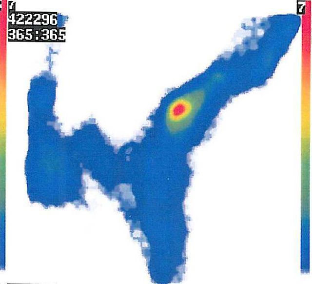
In equine, enostosis-like lesions were first described as multifocal sclerotic areas within the medullary cavity of long bones and were reported in six Thoroughbreds, three Standardbreds and one Appoloosa(1). Most of the horses had a history of chronic lameness. The bone sclerotic areas seen on radiographs and as abnormal increased radioisotope uptake at scintigraphic examination were localized to the diaphyseal areas of the long bones. The lesions were endosteal and/or medullary. These lesions were named enostosis-like in order to differentiate them from enostosis (bone islands seen in man) and panosteitis seen in dog. Later, Ramzan (4) also described enostosis-like lesions in 12 Thoroughbreds. Some of these lesions appeared related to lameness, but in many horses no association could be made. The author concluded that enostosis-like lesions are transient phenomena, with scintigraphic and radiographic resolution occurring over months. Multiple enostosis-like lesions has been reported in a racing Thoroughbred.(3) As treatment of the disease, box rest and controlled exercise during 1-6 months have been suggested. Most of the horses recover and return to working or racing.
The etiology and pathogenesis of theses lesions are not known. Often the bone lesions have been found in the diaphyseal area close to the nutrient foramen, hence bone infarcts has been discussed as a cause. Stress fractures are also mentioned, but never proven to be associated with the radiodense areas. Microscopic examinations of the enostosis-like lesions in horses are not presented in the literature, hence a comparison of the present case and the reported enostosis-like lesions can not be made; however, the radiographic and scintigraphic findings are similar.
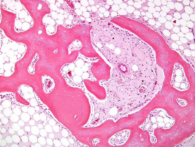
The present microscopic picture is that of a non-neoplastic non-specific bone growth with osteoblast activity, within the medullary cavities, very similar to changes seen microscopically in canine panosteitis. â¨This horse had had a long history of gait asymmetries also involving tendonitis and the owner elected euthanasia. The enostosis-like lesions probably would have resolved and was not the reason for euthanasia.
JPC Diagnosis:
Conference Comment:
Clinically, an increase in radiodensity in the marrow is initially seen and is caused by rapidly expanding areas of fibrovascular tissue that is quickly remodeled and converted to woven bone. Because of this rapid cyclical process of formation and resorption of bone, both osteoblasts and osteoclasts are often observed in the same microscopic field. Resting and reversal lines can also be present in relatively close proximity within medullary trabecular bone.(5)
Inflammation is usually not a feature of this disease, and the lesions resolve spontaneously over time. The cause is currently unknown, but canine distemper virus has been suggested as a potential suspect.(5)
References:
2. Gould CF, Ly JQ, Lattin Jr GE, Beall DP, Sutcliffe JB: Bone tumor mimics: avoiding misdiagnosis. Curr Probl Diagn Radiol 36:124-141, 2007
3. Jones E, McDiarmid A: Case report. Multiple enostosis-like lesions in a racing Thoroughbred. Equine Vet Education. 17:92-95, 2005
4. Ramzan PHL: Equine enostosis-like lesions: 12 cases. Equine Vet J. 14:143-148, 2002
5. Thompson K: Diseases of bones and joints. In: Pathology of Domestic Animals, ed. Maxie G, 5th ed., pp.104-105, vol 1. WB Saunders, Edinburgh, Scotland, 2007