Signalment:
Five-year-old, captive hatched, male tentacled snake (
Erpeton tentaculatum)One of a group of tentacled snakes with a 3 or 4 month history of skin lesions. Six snakes died despite treatment.
Gross Description:
Numerous 2-4 mm diameter irregular slightly depressed rough tan lesions are scattered in the skin of the entire body.
Histopathologic Description:
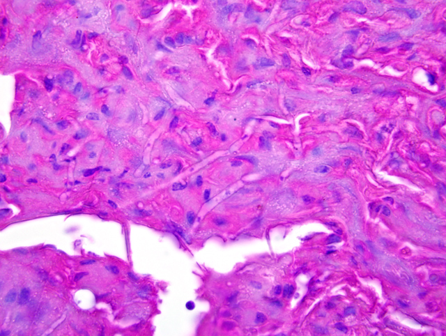
Multifocally, there is full-thickness epidermal necrosis with invasion and expansion of the necrotic tissue by numerous branching, septate fungal hyphae (
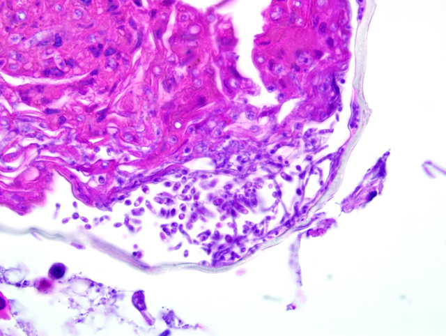
Fig. 4-1) and rare arthroconidia (
Fig. 4-2). In some affected areas, myriads of mixed bacteria are also present. Heterophils multifocally infiltrate the epidermis and dermis and are especially abundant in the dermis in areas where there is separation or loss (ulceration) of the necrotic epithelium. Less affected epidermis is diffusely mildly hyperplastic. Spongiosis and cytoplasmic vacuolation are common.
Morphologic Diagnosis:
Skin: Severe multifocal ulcerative and necrotizing dermatitis with intralesional fungi (etiology:
Chrysosprorium anamorph of
Nannizziopsis vriesii)
Lab Results:
Fungal culture of skin grew
Chrysosprorium anamorph of
Nannizziopsis vriesii
Condition:
Chrysosprorium anamorph of Nannizziopsis vriesii
Contributor Comment:
The
Chrysosporium anamorph of
Nannizziopsis vriesii (CANV) has relatively recently been recognized as a pathogenic fungus that can cause significant morbidity and mortality in some species of reptiles.(2,4,6) In previous reports, dermatomycoses in snakes and lizards have been ascribed to several different species of fungi including dermatophytes such as
Geotrichium sp.,
Trichosporon sp., and numerous other soil fungi (e.g.,
Aspergillus, Candida, Cladosporium, Fusarium, and
Mucor).(2,4,6) It is proposed that in many of these cases the fungus was misidentified and infection was due to the CANV.(2,4)
Nannizziopsis vriesii is a sexually reproductive keratinophilic species in the phylum Ascomycota, order Onygenales, that was originally isolated from the tissues of a lizard (
Ameiva sp.).(4,6) Sexual fruiting bodies are formed on nutrient-poor medium at 30 degrees C. The asexual (mitotic) state seen in histologic lesions is typical of
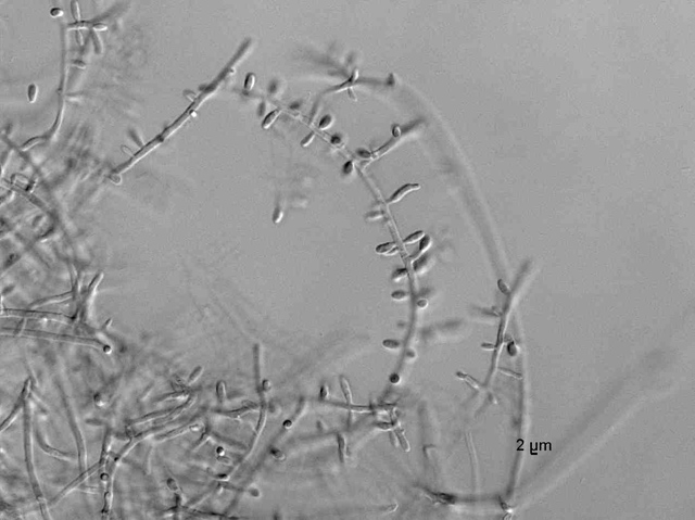
Chrysosporium and produces solitary conidia (aleurioconidia) and arthroconidia. Although culture is necessary to definitively identify the CANV from lesions in affected reptiles, the characteristic appearance of the arthroconidia in histologic section is a key diagnostic feature. The CANV was cultured from the skin of the snake submitted for this conference (Fig. 1). Arthroconidia were not seen histologically in this snake but were seen in the skin lesions of other snakes in this group (Fig. 2). The origin of infection with the CANV in this and other cases is unknown. An extensive survey of skin samples from healthy reptiles did not recover any of the CANV.(7) Thus,
N. vriesii does not appear to be a common constituent of the microflora of healthy reptiles. An environmental source is postulated but has not been demonstrated. Factors that might predispose a reptile to infection and disease are also not known.
JPC Diagnosis:
Scaled skin: Epidermitis, necrotizing and ulcerative, multifocal, marked with intralesional fungi
Conference Comment:
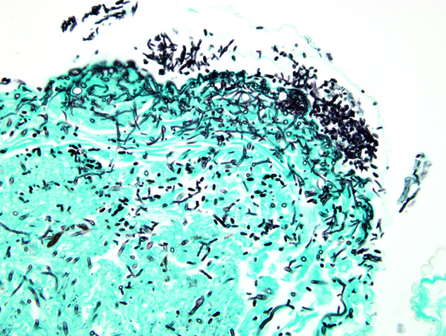
Within the last 10 years, CANV has been identified as the causative agent of dermatomycosis in several reptilian species to include green iguanas, bearded dragons, brown tree snakes, a salt-water crocodile, and veiled chameleons as well as tentacled snakes.(1,2,3,4,5,8) A necrotizing and granulomatous dermatitis is commonly seen in these species with some species variation.(1,2,3,4,5,8) One of the bearded dragons infected with CANV also had a granulomatous hepatitis with intralesional hyphae.(3) Histologically, CANV is found in necrotic lesions and appears as hyaline, septate, branching hyphae often 2-4 um in width with characteristic arthroconidia.(2)
In the case of the tentacled snakes, it was speculated by the author of the article that failure to maintain an acidic environment predisposed these snakes to skin infection. These snakes normally inhabit slow moving acidic streams at a pH of 6-6.5, and the affected snakes were kept in water at a pH of > 8.(2) Snakes kept in water at a pH of 7 did not develop lesions.(2)
The morphologic features of CANV in tissue section are best demonstrated with special stains such as GMS (
Fig. 4-3). The contributor submitted a superb image of hyphae and arthroconidia growing in culture (
Fig. 4-4).Â
References:
1. Abarca ML, Martorell J, Castellia G, Ramis A, Cabanes FJ: Cutaneous hyalohyphomycosis caused by
Bhrysosporiuim species related to
Nannizziopsis vriesii in two green iguanas (Iguana iguana). Med Mycol.Â
46(4):349-354, 2008
2. Bertelsen MF, Crawshaw GJ, Sigler L, Smith DA: Fatal cutaneous mycosis in tentacled snakes (
Erpeton tentaculatum) caused by the
Chrysosporium anamorph of
Nannizziopsis vriesii. J Zoo Wild Med
36(1): 82-87, 2005
3. Bowman MR, Pare JA, Sigler L, Naeser JP, Sladky KK, Hanley CS, Helmer P, Phillips LA, Brower A, Porter R: Deep fungal dermatitis in three inland bearded dragons (
Pogona vitticeps) caused by the
Chrysosporium anamorph of
Nannizziopsis vriesii. Med Mycol.Â
45(4) 371-376, 2007
4. Nichols DK, Weyant RS, Lamirande EW, Sigler L, Mason RT: Fatal mycotic dermatitis in captive brown tree snakes (
Boiga irregularis). J Zoo Wildl Med
30(1): 111-118, 1999
5. Pare A, Coyle KA, Sigler L, Maas AK, Mitchell RL: Pathogenicity of the
Chrysosporium anamorph of
Nannizziopsis vriesii for veiled chameleons (
Chamaeleo calyptratus). Med Mycol.Â
44(1):25-31, 2006
6. Par+�-� JA, Sigler L, Hunter DB, Summerbell RC, Smith DA, Machin KL: Cutaneous mycoses in chameleons caused by the
Chrysosporium anamorph of
Nannizziopsis vriesii (Apinis) Currah. J Zoo Wildl Med
28(4): 443-453, 1997
7. Par+�-� JA, Sigler L, Rypien K, Gibas CF, Hoffman TL: Cutaneous fungal microflora of healthy squamate reptiles and prevalence of the
Chrysosporium anamorph of
Nannizziopsis vriesii: Proc AAZV, AAWV, ARAV NAZWV Joint Conf. pp. 36-38, 2001
8. Thomas AD, Sigler L, Peucker S, Norton JH, Nielan A:
Chrysosporium anamorph of
Nannizziopsis vriesiii associated with fatal cutaneous mycoses in the salt-water crocodile (
Crocodylus porosus). Med Mycol.Â
40(2):143-151, 2002