Signalment:
Gross Description:
Histopathologic Description:
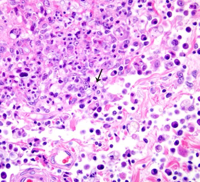
Sections of pancreas (not submitted) contained multifocal to coalescing areas of necrosis with predominate histiocytes and occasional plasma cells, lymphocytes and neutrophils. Round to oval and angular, 5-15 microns, colorless, refractile organisms were phagocytized in macrophages and free within these necrogranulomatous aggregates.
Sections of lung (not submitted) contained multifocal areas of alveolar collapse and interstitial thickening with neutrophils, macrophages, lymphocytes and plasma cells.Â
Morphologic Diagnosis:
(Following tissues not included in submitted histologic sections) 2. Pancreas: Moderate to severe, multifocal to coalescing, necrogranulomatous pancreatitis with intralesional organisms consistent with Prototheca sp. 3. Lung: Moderate, multifocal, pyogranulomatous interstitial pneumonia.
Condition:
Contributor Comment:
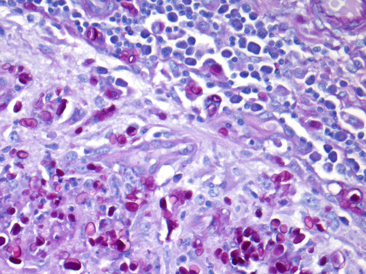
Prototheca sp. are round, oval, or angular cells that are 8-20 micrometers in diameter, have a refractile wall and contain granular cytoplasm. The organism reproduces by endosporulation and can be seen histologically as morula of 2-20 daughter cells within a single organism. The morula ruptures and releases the individual daughter cells. The cell wall of Prototheca stains poorly with hematoxylin-and-eosin, but stains strongly positive to stains for carbohydrate, such as PAS, Gridley, Bauer, and GMS.Â
Prototheca sp. resembles Chlorella sp., but can be distinguished by PAS-positive starch granules in the cytoplasm of Chlorella. In fresh smears of lesions that contain Chlorella, the organisms are green due to the presence of chlorophyll.2
JPC Diagnosis:
Conference Comment:
Infection is thought to occur through traumatic inoculation, ingestion, or wound contamination. The immune status of an individual appears to play a role in acquiring the infection as well as lesion extent and distribution, although infections have been found in both immunocompetent and immunosuppressed patients.4
Clinical manifestation of protothecosis in mammals is to a certain extent species specific. In cattle, mastitis due to P. zopfii is the most common presentation of protothecosis in cattle1,4 with occasional spread into adjacent lymph nodes, while protothecosis in the dog, also primarily due to P. zopfii4, is principally a systemic disease with multi-organ involvement and a predilection for the eyes and brain.2,3 Protothecosis in cats, due to P. wickerhamii, localizes in the skin, and may be successfully treated by wide surgical excision.3,4
In dogs the most consistent presenting clinical sign of systemic protothecosis is hemorrhagic colitis.4 In fact, the colon and the rectum appear to be primary sites of replication even without clinical evidence of colitis.4 A proposed pathogenesis includes initial colonization of the colonic mucosa following ingestion of large numbers of infective organisms, followed by penetration of the gut wall and systemic spread via blood vessels and lymphatics.4
Table extracted from Stenner et al.4
| - | Shape | Sporangia | Sporangiospores |
| Prototheca zopfii | oval or cylindrical | 14-25 um | up to 20 |
| Prototheca wickerhamii | round | 7-13 um | up to 50 |
References:
2. Jones T, Hunt R, King N: Diseases caused by fungi. In: Veterinary Pathology, 6th ed., pp. 534-535. Williams & Wilkins, Baltimore, MD, 1997
3. Schultze AE, Ring RD, Morgan RV, Patton CS: Clinical, cytologic and histopathologic manifestations of protothecosis in two dogs. Vet Ophthalmol 1:239-243, 1998
4. Stenner VJ, Mackay B, King T, Barrs VR, Irwin P, Abraham L, Swift N, Langer N, Bernays M, Hampson E, Martin P, Krockenberger MB, Bosward K, Latter M, Malik R: Protothecosis in 17 Australian dogs and a review of the canine literature. Med Mycol 45:249-266, 2007