Signalment:
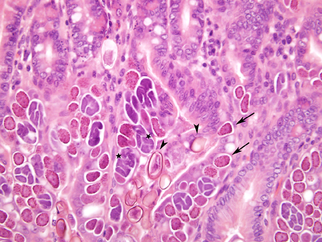
Histopathologic Description:
Morphologic Diagnosis:
Condition:
Contributor Comment:
Despite the striking degree of mucosal involvement, no diarrhea was reported, and no evidence of it was noted at necropsy. Nonetheless, E. magna is reported to be of moderate pathogenicity4.Â
Intestinal coccidiosis continues to be common in rabbits, probably due to multiple factors, including the frequency of inapparent infections, the massive number of oocysts shed by infected rabbits, and the resistance of oocysts to many disinfectants. More than 12 species of intestinal coccidia have been reported in rabbits 6, most of which live in the small intestine. Definitive diagnosis requires examination of sporulated oocysts. In our experience, oocysts will sporulate in fecal samples left at room temperature (presumably as in the wild) or in the refrigerator for several days, but sporulation is more reliably accomplished by incubation at room temperature for 1-5 days in a potassium dichromate solution. Sporulated oocysts are readily speciated based on size, appearance, and the appearance, if present, of the micropyle and residual body.
The life cycle of E. magna is typical of Eimeria spp. All Eimeria are host-specific and have a direct life cycle2. Oocysts are not infective until sporulation, so ingestion of cecotroph feces does not result in autoinfection. Ingestion of sporulated oocysts (sporocysts) results in release (excystation) of sporozoites. These invade enterocytes, round up and form trophozoites, and multiply asexually by schizogony (merogony), forming schizonts (meronts) that may contain more than 100,000 merozoites. Merozoites escape the host cell, resulting in death of that cell. Each merozoite can then invade another host cell for the next generation. The number of asexual generations is characteristic of each Eimeria sp., E. magna has been variously reported as having four or five3, 5 asexual generations prior to gametogony. In gametogony, the final generation merozoites form either macrogametocytes (female) or microgametocytes (male). Macrogametocytes have a single nucleus, and numerous peripheral PAS-positive granules. Microgametocytes are multinucleate. Each nucleus becomes incorporated into a small biflagellate sperm-like microgametocye. After fertilization by the microgametocyte, the macrogametocyte develops into an oocyst. It has been estimated that one oocyst of E. magna can produce more than 25,000,000 oocysts in a susceptible host4. Given that feces from asymptomatic rabbits may contain more than 400,000 oocysts/gram, the potential for massive infection is apparent. Clinical disease is thought to result primarily from loss of functional mucosa and loss of mucosal barrier integrity as cells are lost.
We personally find it interesting that such massive infection did not cause significant debility (note the normal mesenteric fat) or diarrhea. The general sparing of the crypts may indicate that the rabbit retains sufficient epithelial replacement capacity, although no increase in mitotic rate was noted. Sparing of the crypts and the observation of generally less developed forms deeper along the villus also suggest two evolutionary adaptations of this parasite: first, that sparing the crypts is advantageous to the replication of the parasite as the host is not rapidly killed; and second, that invasion of cells near the crypt or along the side of the villus may be preferable as those cells are less likely to be shed prior to completion of a particular phase in the life cycle.Â
JPC Diagnosis:
Conference Comment:
| Eimeria and Isospora of Animals | ||
| Â Â | ||
| Geese & ducks | E. truncata | Kidney |
| Sandhill whooping cranes | E. reichenowi | Disseminated |
| Parrots | E. psittaculae | Intestine |
| Chicken | E. acervulina | Duodenum |
| Chicken | E. necatrix | Mid-intestine |
| Chicken | E. tenella | Ceca |
| Â Â | ||
| Cattle | E. bovis | Small intestine, cecum, colon |
| Â Â | ||
| Sheep | E. ashata | Small intestine |
|  | E. bakuensis | Small intestine |
|  | E. ovinoidalis | Ileum, large intestine |
| Â Â | ||
| Goats | E. Christenseni | Small intestine |
|  | E. arlongi | Small intestine |
|  | E. ninakohlyakimovea | Large intestine |
| Â Â | ||
| Horses | E. leukarti | Small intestine |
| Â Â | ||
| Swine | I. suis | Intestine |
|  | E. debliecki |  |
|  | E. porci |  |
|  | E. scabra |  |
| Â Â | ||
| Dogs | I. canis | Ileum, cecum occasionally |
| Â Â | ||
| Cats | I. felis | Small intestine, colon occasionally |
| Â Â | ||
| Mice | E. falciformis | Colon |
| Â Â | ||
| Rabbit | E. stiedae | Bile ducts |
|  | E. intestinalis | Ileum, cecum |
|  | E. flavescens | Ileum, cecum |
| Â Â | ||
| Guinea pig | E. caviae | Large intestine |
| Â Â | ||
| Ferret | E. furonis | Gallbladder, bile duct |
Conference participants briefly reviewed the coccidian life cycle. Oocysts are shed in feces and sporulate. The oocysts of each species are morphologically distinct, but share similar features. The oocysts of Eimeria have four sporocysts, each with two sporozoites, with a total of eight sporozoites in each oocyst. The oocysts of Isopora have two sporocysts, each with four sporozoites, with a total of eight sporozoites in each oocyst. Ingested sporozoites excyst in the intestine and invade epithelial cells where they round up and form trophozoites. Asexual replication or schizogeny follows forming schizonts containing merozoites. The schizonts rupture, releasing the merozoites, which infect other epithelial cells and continue to replicate. Merozoites eventually form sexual stages (male-microgamete, female-macrogamete) which unite to form oocysts.2
Conference attendees also reviewed the ultrastructural features of apicomplexans, specifically Toxoplasma: parasitophorous vacuole, rhoptries, micronemes, apical conoid, apicoplasts, and dense granules.Â
This case was reviewed in consultation with Dr. Chris Gardiner, AFIP consultant in veterinary parasitology. We are grateful to Dr. Gardiner for his comments and advice on this interesting case.
References:
2. Pakandl M, Eid AN, Licois D, Coudert P: Eimeria magna Perard, 1925: study of the endogenous development of parental and precocious strains. Vet Parasitol 1996, 65: 213-222.
3. Percy DH, Barthold SW: Pathology of Laboratory Rodents and Rabbits. Ames, Iowa State University Press, 2001.
4. Ryley JF, Robinson TE: Life cycle studies with Eimeria magna Perard, 1925. Z Parasitenkd 1976, 50: 257-275
5. Schoeb TR, Cartner SC, Baker RA, Gerrity LW: Parasites of rabbits. Flynn's Parasites of Laboratory Animals. Edited by Baker DG. Blackwell Publishing, 2007, pp. 451-499.