WSC 22-23
Conference 20
Case I:
Signalment:
14-week-old, male, Saanan goat
History:
One of five kids from a herd of 60 goats that developed progressive onset hindlimb ataxia and sternal recumbency terminally.
Gross Pathology:
No specific findings. CNS appeared normal.
Laboratory Results:
Liver copper 0.01 mmol/kq, (normal range 0.06 - 2.5 mmol/kg - values for sheep)
Liver molybdenum 9.54 µmol/kq,(normal range 15.6 - 62.0 µmol/kg - values for sheep)
Microscopic Description:
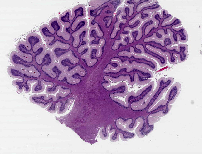
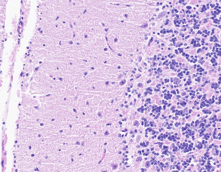
Cerebellum: multifocally, Purkinje cells are lost, and a small number are shrunken/angulated with cytoplasmic eosinophilia and karyopyknosis. Ectopic Purkinje cells noted within an irregularly thinned granular layer. Concurrent prominence of reactive Bergmann glial cells. Occasional light perivascular cuffs of admixed lymphocytes and plasmacytes noted in cerebrum. No significant changes noted elsewhere in brain or in spinal cord.
Contributor’s Morphologic Diagnoses:
Cerebellum: degeneration and dysplasia (particularly affecting Purkinje cells), multifocal, moderate
Contributor’s Comment:
A diagnosis of delayed enzootic ataxia (swayback) in this goat kid was based on: signalment, clinical signs, the microscopically visible cerebellar lesions and low liver copper concentrations. This neurological disease of sheep and goats is generally accepted to result from a primary deficiency in the intake of copper or a secondary deficiency caused the reduced absorption/availability of copper during pregnancy.3 In the secondary form dietary molybdenum, zinc, iron and cadmium can interfere with copper absorption.1,6 Molybdenum is an important antagonist for copper as (in combination with sulfate), it forms thiomolybdates in the rumen which can complex with copper, limiting its absorption.3 All three affected kids had liver copper concentrations below the normal range. There was no history of swayback in this goat herd and the dams’ diet consisted of grass, hay and sheep concentrate (unknown quantity). The goats had access to mineral licks/blocks and were being housed at night.
Clinically, two forms of enzootic ataxia in goat kids and lambs are described: a congenital form, more commonly reported in lambs develops in utero and manifests in the first few days of life; and a delayed form in both lambs and kids up to six months of age.1,2 Affected animals develop progressive neurologic signs such as ataxia (swaying), hindlimb paralysis, and blindness. Lesions are found in the cerebrum, brainstem, and spinal cord in the congenital form, but are limited to the brainstem and cord in the delayed form. Cerebral lesions may be found in approximately 50% of congenitally infected lambs but rarely in kids: these consist of bilaterally symmetrical cavities in the white matter.3,6 Microscopically visible lesions in both white and gray matter regions of the brainstem and spinal cord in both congenital and delayed forms are similar in both goats and sheep. Neurons in the medullary reticular, red and lateral vestibular nuclei in the brainstem and in the lateral and ventral spinal cord exhibit degenerative changes: chromatolysis, cytoplasmic eosinophilia and pyknosis/margination of nuclei. Wallerian-type axonal degeneration is observed in dorsolateral and ventromedial spinal cord funiculi.3,6 Ultrastructural observations in the goat and the particular involvement of neurons having long processes within the CNS or extending into peripheral nerves, suggest that the neuraxon rather than its myelin sheath is targeted in delayed enzootic ataxia.14 While myelin degradation in this eventuality would then be expected as a secondary event, myelin in this context is qualitatively abnormal and therefore potentially unstable.3 Accompanying astrogliosis is mild.3,6 Cerebellar cortical lesions, particularly targeting Purkinje cells as described here are reported more commonly in goats than in sheep.2,14
Copper is required for the activity of several enzymes essential for neural function, including cytochrome oxidase (mitochondrial respiration) and copper-zinc superoxide dismutase (antioxidant activity). Suggested mechanisms contributing to the development of these lesions include altered function of cytochrome oxidase in mitochondria leading to suppression of mitochondrial respiration and reduced phospholipid synthesis. This energy failure is likely to result in axonal/neuronal degeneration. The generation of excessive reactive oxygen species as a consequence of inadequate function of superoxide dismutase ultimately results in oxidative injury within the CNS.3,6
Copper deficiency is more commonly encountered in grazing animals because of the poor availability of this element in grass. Foodstuffs from which copper is more readily absorbed are those low in fiber such as cereals. Preservation of grass in the form of hay or silage improves copper availability.2 The fact that goats have a lower capacity to store copper in the liver than do sheep10,14 would suggest this species requires a higher daily intake. In previous instances of enzootic ataxia in goats, sheep concentrate, low in copper, had been used as supplementary food. No further cases occurred following the replacement of sheep concentrate by concentrate appropriate for cattle and horses.14
Differential diagnoses in this case would include white muscle disease, spinal trauma/vertebral body abscess, caprine arthritis encephalitis, meningoencephalitis, and possible toxicity (lead, organophosphate). Enzootic ataxia may be prevented through adequate copper supplementation of the dam during the latter half of pregnancy.2
Contributing Institution:
Room 012, Veterinary Sciences Centre, School of Veterinary Medicine, University College Dublin, Belfield, Dublin 4, Ireland
JPC Diagnosis:
Cerebellum: Purkinje and granular cell loss, segmental, moderate.
JPC Comment:
The contributor provides a good overview of the gross lesions, histologic findings, and pathogenic features of this classic entity in small ruminants. Another example of copper deficiency in a young goat was seen in WSC 2021 Conference 2 Case 1. Readers are encouraged to review that case which also demonstrated neuroaxonal degeneration in the spinal cord secondary to copper deficiency. Copper deficiency has also been implicated as a cause of laryngeal neuropathy in adult goats. In a 2017 Veterinary Pathology study, researchers described goats with severe copper deficiency that had both ataxia and stridor, while moderately affected goats had ataxia without stridor.12 The severely affected goats demonstrated atrophy of laryngeal muscles appreciable both grossly and histologically, with Wallerian degeneration of the recurrent laryngeal nerve.12 Interestingly, both sets of goats also had increased serum iron levels, with the more severely affected goats having higher levels, indicating that high dietary iron may have led to copper deficiency.12
As the contributor describes, copper is an essential cofactor in the activity of many enzymes in the body. Copper is an essential component of tyrosinase, which is involved in the rate-limiting step in melanin synthesis, and deficiency results in depigmentation of the hair or wool, causing a rusty brown discoloration and the formation of spectacles in the fur around the eyes (periocular achromotrichia).5,13 Copper is also important for oxidation of sulfhydryl groups during keratinization, and deficiency causes hair shafts in sheep to be straighter with less crimp (“string” or “steely” wool).5,13 Alterations in coat color and texture due to copper deficiency have been reported in dogs, cattle, sheep, and moose.5
Copper is also a component of lysyl oxidase and is required for the proper crosslinking of collagen and elastin.4 Copper deficiency can lead to fragile bones and cartilage in dogs, lambs, foals, calves, and pigs.4 In dogs, copper deficiency is associated with decreased osteoblast activity and can produce metaphyseal lesions similar to the scorbutic lattice of vitamin C deficiency.4 In foals, copper deficiency may cause the articular epiphyseal cartilage complex to lyse which may resemble osteochondrosis.9
Prolonged copper deficiency has been associated with sudden death and myocardial scarring in cattle in Australia and Florida (“falling disease”).11 In a recent study on the effect of prolonged copper deficiency on cardiac function, researchers found histologically-apparent fibrosis in the ventricular myocardium, severe decrease in copper-zinc superoxide dismutase activity, and significantly increased levels of lipid peroxidation byproducts.8
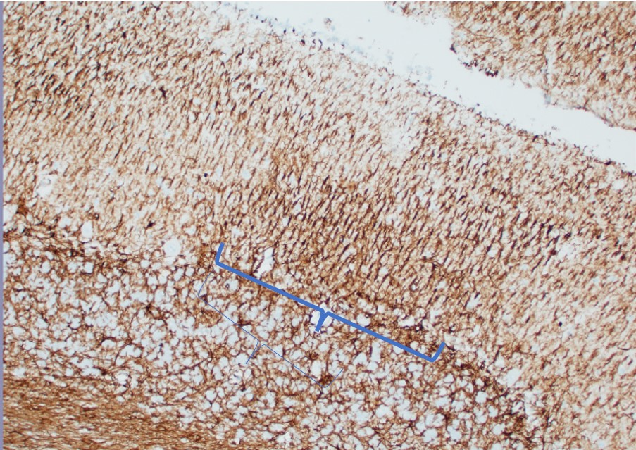
This week’s moderator, LTC Keith Koistinen, described the function of Bergmann glial cells (specialized radial glia astrocytes) which are prominent in this case: to provide scaffolding for the migration of Purkinje cells. Bergmann glial cells are difficult to distinguish solely on H&E. In this case, the molecular layer looks slightly more dense in the areas of Purkinje cell loss, suggesting proliferation of Bergmann glial cells; however, definitive identification of Bergmann glial cells requires GFAP immunohistochemistry. The moderator and participants also remarked on the fact that Purkinje cells were present in the granular layer, reflective of defective migration referenced in the contributor’s morphologic diagnosis of Purkinje cell dysplasia. The moderator also described that the best tissue for submission for copper analysis is brains for neonates and liver for adults.
References:
- Allen AL, Goupil BA, Valentine BA. A retrospective study of spinal cord lesions in goats submitted to 3 veterinary diagnostic laboratories. Can Vet J. 2012;53:639-642.
- Banton MI, Lozano-Alarcon F, Nicholson SS, Jowett PLH, Fletcher J, Olcottet BM. Enzootic ataxia in Louisiana goat kids. J Vet Diagn Invest. 1990;2:70-73.
- Cantile C, Youssef S. Nervous system. In: Maxie MG, ed. Jubb, Kennedy and Palmer’s Pathology of Domestic Animals. 6th 1. St. Louis, MO:Elsevier Ltd; 2016:328-329.
- Craig LE, Dittmer KE, Thompson KG. In: Maxie MG, ed. Jubb, Kennedy and Palmer’s Pathology of Domestic Animals. 6th 1. St. Louis, MO:Elsevier Ltd; 2016:66.
- Mauldin EA, Peters-Kennedy J. Integumentary System. In: Maxie MG, ed. Jubb, Kennedy and Palmer’s Pathology of Domestic Animals. 6th 1. St. Louis, MO:Elsevier Ltd; 2016:558.
- Miller AD, Zachary JF. Copper deficiency. In: Zachary JF, ed. Pathologic Basis of Veterinary Disease. 6th St. Louis, MO: Elsevier Ltd; 2017:887-888.
- Miller AD, Porter BF. Nervous system. In: Zachary JF, ed. Pathologic Basis of Veterinary Disease. 7th ed. St. Louis, MO: Elsevier; 2022:973-974.
- Olivares RWI, Postma GC, Schapira A, et al. Biochemical and Morphological Alterations in Hearts of Copper-Deficient Bovines. Biol Trace Elem Res.2019; 189(2):447-455.
- Olson EJ, Dykxtra JA, Armstrong AR, Carlson CS. Bones, Joints, Tendons, and Ligaments. In: Zachary JF, ed. Pathologic Basis of Veterinary Disease. 7th ed. St. Louis, MO: Elsevier; 2022:1060.
- Papachristodoulou C, Stamoulis K, Tsakos P, Vougidou C, Vozikis V, Papadopoulou C, Ioannides, K. Liver concentrations of copper, zinc, iron and molybdenum in sheep and goats from northern Greece, determined by energy-dispersive x-ray fluorescence spectrometry. Bull Environ Contam Toxicol 2015; 94:460-467.
- Robinson WF, Robinson NA. Cardiovascular System. In: Maxie MG, ed. Jubb, Kennedy and Palmer’s Pathology of Domestic Animals. 6th 1. St. Louis, MO:Elsevier Ltd; 2016:34.
- Sousa RFA, Almeida VM, Neto JE, et al. Laryngeal Neuropathy in Adult Goats with Copper Deficiency. Vet Pathol. 2017; 54(4): 676-682.
- Welle MM, Linder KE. The Integument. In: Zachary JF, ed. Pathologic Basis of Veterinary Disease. 7th ed. St. Louis, MO: Elsevier; 2022:1201.
- Wouda W, Borst GHA, Gruys E. Delayed swayback in goat kids, a study of 23 cases, Vet Q. 1986; 8 (1):45-56.