CASE 3: MSU Case#1 (4065133-00)
Signalment: 3.5-year-old, male, white-tailed deer, Odocoileus virginianus
History:
This buck was harvested by a hunter during rifle season. He submitted the deer for examination because of skin lesions.
Gross Pathology:
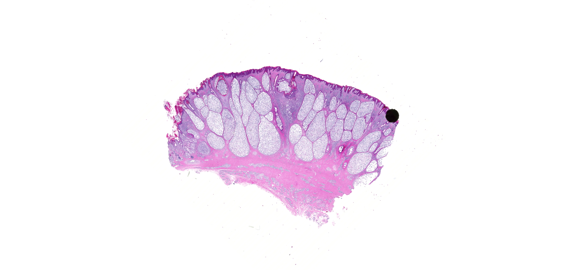
The skin over the muzzle, face, ears, and limbs had patchy areas of thickening, alopecia, and crusts. On cross-sections of affected skin, numerous round to elongate, pale, 2-5mm diameter nodules were visible. In addition, the subcutaneous tissues of the muzzle were diffusely swollen and edematous.
Laboratory results:
PCR testing for BVD on spleen was negative. PCR on pooled lung, liver and spleen for Epizootic Hemorrhagic Disease virus was negative. Immunohistochemical staining of obex and retropharyngeal lymph nodes for Chronic Wasting Disease prion protein was negative.
Microscopic description:
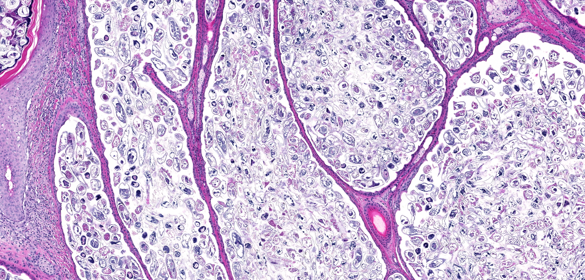
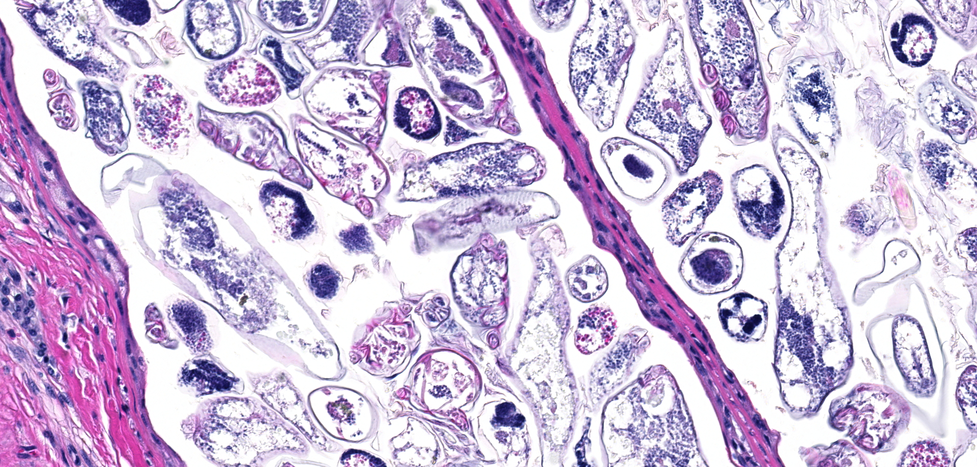
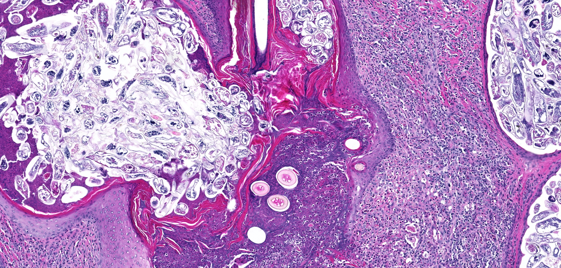
Sections of skin contained markedly ectatic hair follicles filled with myriads of cigar-shaped arthropod parasite adults, nymphs, larva and eggs. Adults measured 140-180µm long, 25-40µm diameter, with 4 pairs of stubby legs, and contained striated muscles internally. Follicles exhibited loss of hair shafts, moderate hyperkeratosis, secondary bacterial colonization, with predominantly lymphoplasmacytic folliculitis. Some hair follicles had ruptured resulting in granulomatous to pyogranulomatous furunculosis associated with mites outside of the hair follicles. Overlying epidermis was moderately thickened, with rete peg formation, orthokeratotic hyperkeratosis, and variable amounts of serocellular crusting. The dermis was expanded by mixed lymphoplasmacytic, eosinophilic, and neutrophilic infiltrates.
Contributor's morphologic diagnosis:
Skin: severe follicular ectasia, with lymphoplasmacytic folliculitis and granulomatous furunculosis, and numerous intra-follicular arthropod mites consistent with Demodex sp.
Contributor's comment:
Demodicosis is a fairly straightforward histologic diagnosis in most severely affected individuals in many different species of animal. The species of demodectic mite most commonly reported in white-tailed deer is Demodex odocoilei. Other species of deer known to be affected by this parasite include mule deer and black-tailed deer.3,5 European deer species including red deer, roe deer, Sambar deer and sika deer are reported to be infested with Demodex kutzeri.3 While the mites in this report are in the right size range reported for nymphs of Demodex odocoilei, no specific molecular identification was performed.3
The degree of mite infestation in this case is impressive. While I have examined a number of deer cases with demodicosis, I have never encountered one with grossly visible ectatic follicles, although other authors have reported these nodules.5 I would estimate that there were several hundred organisms per infected hair follicle. One recent paper cites an estimate of 6.3 mites per hair follicle. Another unusual feature was the grossly swollen edematous muzzle in this animal. My colleagues in the MI Department of Natural Resources have seen this several times and refer to this as the "Bullwinkle J. Moose Syndrome", since the muzzle appears bulbous and swollen as naturally occurs in moose. I know of no reported explanation for this syndrome, nor of its occurrence in any other mammalian species in association with demodicosis infestations. Possible pathogeneses may include secondary bacterial infection of the follicles and skin leading to a local bacterial toxin release, or follicular ectasia and granulomatous furunculosis leading to physical obstruction of lymphatic drainage causing secondary subcutaneous edema.
The severe degree of pathology associated with this case is atypical for demodicosis. Therefore, we considered underlying immunosuppression as occurs in some cases of generalized canine demodicosis. However, fecal examination failed to reveal heavy gastrointestinal parasitism, examination of lymph nodes and lungs revealed no gross or microscopic evidence of bovine tuberculosis (endemic in MI deer), and testing for BVD, CWD and EHD were all negative. Furthermore, the deer was not in poor body condition. However, similar to previous reports, this deer was a male, was harvested in the fall and that time period coincides with breeding season and increases in hormones such as testosterone and cortisol, which may have increased this individual's susceptibility to developing clinical demodicosis.5
Contributing Institution:
Department of Pathobiology
College of Veterinary Medicine
Michigan State University
JPC diagnosis:
Haired skin: Follicular ectasia, diffuse, severe, with multifocal lymphohistiocytic dermatitis, folliculitis, and furunculosis, and myriad intrafollicular mite adults, nymphs, and eggs, white-tailed deer, cervid.
JPC comment:
During conference discussion, there was debate in how best to represent the processes taking place in this tissue. While the furunculosis was a granulomatous process, the dermatitis and folliculitis were of a predominately lymphocytic nature. It was also noted that some slides had focal sarcocysts in the subjacent musculature.
The first reported case of Demodex sp. in the white-tailed deer was in the 1960's, in Georgia, though speciation was not performed. However, a novel Demodex species of white-tailed deer was first described in detail in 1974, including detailed measurements, images of all life stages, and descriptions of several pathogenic manifestations of disease. This novel species was named Demodex odocoilei, and as previously described by the contributor, has been a common finding in white-tailed deer. This is one of a small subset of Demodex species that has been found invading local blood vessels.1,2
While usually host-specific, species of Demodex have been found on different species, including Demodex kutzeri on white-tailed deer, and Demodex odocoilei in Columbian black-tailed deer (Odocoileus hemionus columbianus) and mule deer (Odocoileus hemionus hemionus). A third species of Demodex affecting white-tailed deer was detailed in 2013, with physical parameters and phylogenetic analysis distinguishing it from D. odocoilei. In cases with this new Demodex species, there were multiple 1-2 cm tan cutaneous nodules on the head, legs, and lateral thorax and abdomen, but without alopecia. Histologically, nodules were determined to contain several hundreds to thousands of intrafollicular Demodex mites. There was mild perifollicular lymphocytic and histiocytic dermatitis, but significant inflammation was only present in cases of furunculosis.7
Because Demodex spp are generally thought to be species-host specific, it is curious that we find Demodex species that exist on different host species. Phylogenetic analysis estimates that Demodex kutzeri from elk and D. folliculorum from humans had a common ancestor approximately 85 million years ago. This coincides with the divergence of the two host species, with their common ancestor diverging approximately 84 million years ago. However, when morphologically similar D. kutzeri from elk and mule deer were compared, they had extremely low levels of sequence divergence, suggesting a single species. However, elk and mule deer diverged from each other approximately 7 million years ago. Very few genetic sequences of Demodex have been completed, but in the future may help determine 1) previously unknown species of Demodex, or 2) mechanisms by which they perform host-switching.6
References:
1. Carpenter JW, Freeny JC, Patton CS. Occurrence of Demodex Owen 1843 on a white-tailed deer from Oklahoma. Journal of Wildlife Diseases. 1972;8:112-114.
2. Desch CE, Nutting WB. Demodex odocoilei sp. nov. from the white-tailed deer, Odocoileus virginianus. Can. J. Zool. 1974;52:785-789.
3. Gentes ML, Proctor H, Wobeser G. Demodicosis in a mule deer (Odocoileus hemiones hemiones) from Saskatchewan, Canada. J Wildl Dis 2007;43:758-761.
4. Howerth EW, Nemeth NM, Ryser-Degiorgis MP. Cervidae. In: eds. Terio KA, McAloose D, St. Leger J. Pathology of Wildlife and Zoo Animals. Elsevier: San Diego, CA. 2018:174.
5. Nemeth NM, Ruder MG, Gerhold RW, et al. Demodectic mange, dermatophilosis, and other parasitic and bacteriologic diseases in free-ranging white-tailed deer (Odocoileus virginianus) in the United States from 1975 to 2012. Vet Pathol 2013;51:633-640.
6. Palopoli MF, Tra V, Matoin K, Mac PD. Evolution of host range in the follicle mite Demodex kutzeri. Parasitology. 2017;144(5):594-600.
7. Yabsley MJ, Clay SE, Gibbs SEJ, Cunningham MW, Austel MG. Morphologic and molecular characterization of a Demodex (Acari: Demodicidae) species from white-tailed deer (Odocoileus virginianus). ISRN Parasitology. 2013;342918.