Signalment:
13-year-old, female, Thoroughbred horse (
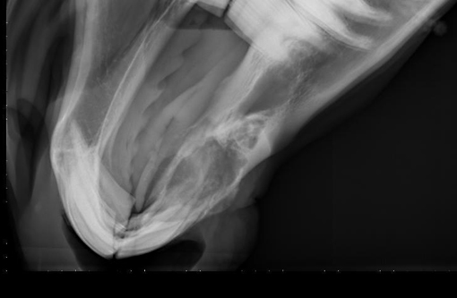
Equus caballus).A rostral mandibular mass on the right side was noted in July 2009. Biopsy samples were non-diagnostic, showing small fragments of new bone formation with a uniform spindle cell population that may have been consistent with reactive new bone or neoplasia, though the cells were not overtly malignant. Radiographs showed an aggressive, lytic, bony lesion, and neoplasia was considered a likely differential. The horse appeared comfortable at this time. She was euthanized eight months later when the mass was perceived to be painful and she had difficulty prehending food.
Gross Description:
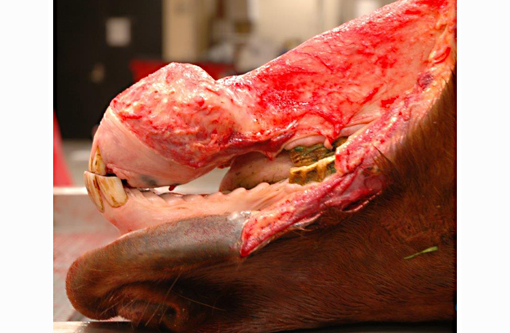
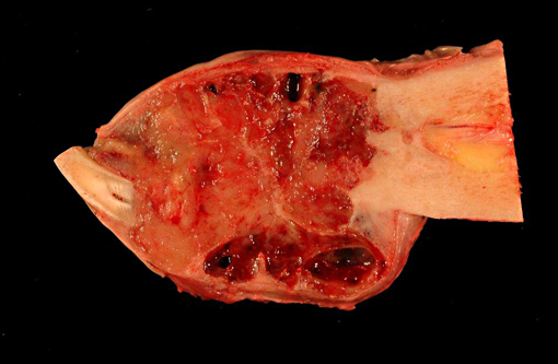
The right rostral mandible is expanded by an 8 x 8 cm, well-demarcated, spherical mass with a smooth but slightly irregular surface. Cut surface of the mass is composed of firm, tan tissue with multifocal regions of cavitation (often with hemorrhage), bone lysis and mineralization. The right mandibular incisors are loose. There is a sharp demarcation between the mass and normal mandibular bone.
Histopathologic Description:
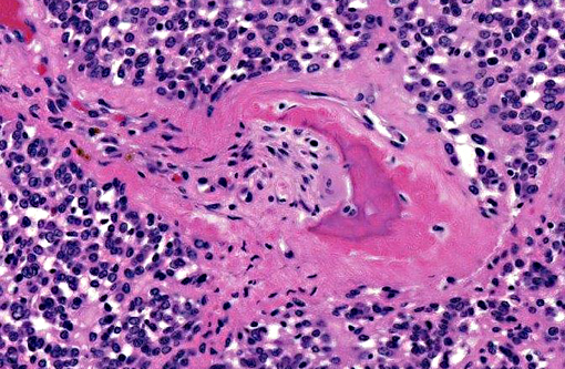
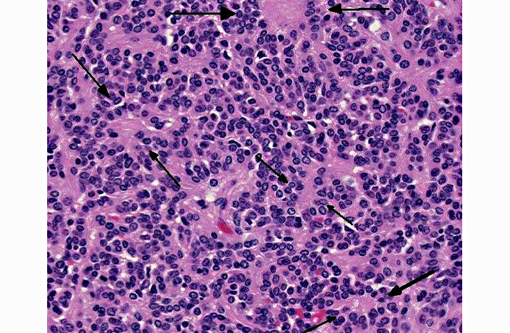
Mandibular mass: Surrounding scant remaining islands of pre-existing mandibular bone is an irregularly multilobulated, densely cellular neoplasm. Lobules are composed of sheets of cells and are separated by moderate fibrovascular stroma. The neoplastic cells are round to polygonal with indistinct cell borders and contain a scant to moderate amount of pale eosinophilic cytoplasm that surrounds a central nucleus. The nuclei are round to oval, and sometimes reniform with coarsely clumped chromatin and rarely have 1-3 small nucleoli. Larger nuclei sometimes have a vesicular appearance to their chromatin. Within the densely cellular lobules, cells are haphazardly arranged with occasional perivascular pseudo-rosettes and rare rosettes (Homer-Wright). There is moderate anisocytosis and anisokaryosis and 6 mitotic figures per 10 high power fields are observed.
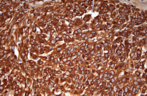
Immunohistochemically, the tumor cells show diffuse, strong staining for glial fibrillary acidic protein (GFAP), and moderate positive reactivity for vimentin and S100. About 60% of the neoplastic cells have weak to moderate cytoplasmic staining for neuron-specific enolase, and about 30% of cells have weak staining for synaptophysin.
Morphologic Diagnosis:
Peripheral primitive neuroectodermal tumor.
Lab Results:
Cultures (aerobic and anaerobic) collected at the initial identification of the mass were negative.
Condition:
Peripheral PNET
Contributor Comment:
Peripheral primitive neuroectodermal tumors (pPNET) are similar to the Ewings sarcoma family of tumors (EFT). In man, there are three main types of tumors in this group including the intraosseous pPNET (Ewings sarcoma), the extraosseous pPNET, and the thoracopulmonary Askins tumor.(3) These tumors are rarely reported in animals, but there is a recent case reported in a camel, a few reports in dogs, and one in a Colobus monkey.(1,4,5) In people, the intraosseous tumors are most common in the long bones, but about 10% of them are reported in the skull. Of the primary tumors in the skull, approximately 10% are in the jaw bones, either the mandible or the maxilla.(3) The recently described camel pPNET had a primary vertebral mass with hepatic metastases.(5)
The pPNETs are often highly aggressive tumors composed of primitive small round cells of putative neuroectodermal phenotype, and generally occur outside the nervous system.(3,5) They often present in nests or sheets of poorly differentiated small round cells with scant cytoplasm and may have rosette or pseudorosette formation. In people, a diagnosis of EFT is strengthened with positive reactivity for MIC2 (CD99) which is not available in veterinary species, and all EFTs in people stain with neuron-specific enolase (NSE).(1,3) Variable staining for synaptophysin and S-100 has been reported in human pPNETs.
Immunohistochemistry in the few reported veterinary cases of pPNET has been somewhat inconsistent with positive triple neurofilament staining reported in one case, and variably positive or negative staining for vimentin, NSE, synaptophysin, and glial fibrillary acidic protein (GFAP).(1,4,5) The variation in reported immunohistochemistry has been postulated to be due to the variable differentiation of the neural crest progenitor cells (neuronal, ependymal, and glial).(5) Our case of a pPNET in a horse mandible most closely resembles the immunohistochemical pattern described in the recent case report in the camel with positive vimentin, GFAP, and NSE staining.
This case is also interesting because the mass was initially noted approximately eight months prior to euthanasia with no evidence of metastases at the time of necropsy, but these tumors are typically aggressive growths that often metastasize.
JPC Diagnosis:
Bone, mandible: Primitive neuroectodermal tumor.
Conference Comment:
As the contributor states, peripheral primitive neuroectodermal tumors (pPNET) have rarely been reported in horses; another case was recently reported in a two-year-old paint horse gelding with multiple lobulated masses bilaterally filling the scrotal cavities, extending through the inguinal canals and along the peritoneal cavity, and infiltrating both kidneys as well as the liver, spleen, mesenteric lymph nodes, mesentery and diaphragm. Histomorphological and immunohistochemical findings were consistent with a pPNET; the neoplastic cells were positive for synaptophysin, S-100, GFAP, NSE, and neurofilament protein (NFP), but negative for muscle-specific actin. Interestingly, in this gelding there appeared to be no bony involvement, whereas most pPNETs reported in veterinary literature, including the mandibular mass presented here, have significant bony involvement.(2)
References:
1. De Cock HE, Busch MD, Fry MM, Mehl M, Bollen AW, Higgins RJ. A peripheral primitive neuroectodermal tumor with generalized bone metastases in a puppy.
Vet Pathol. 2004;41:437-41.
2. Facemire PR, Facemire LM, Honnold SP. Peripheral primitive neuroectodermal tumor in a two-year-old paint horse.
Vet Diagn Invest. 2012; 24:794.
3. Hadfield MG, Quezado MM, Williams RL, Luo VY. Ewing's family of tumors involving structures related to the central nervous system: a review.
Pediatr Dev Pathol. 2000; 3:203-10.
4. Long PH, Schulman FY, Koestner A, Fix AS, Campbell MK, Cameron KN. Primitive neuroectodermal tumor in a two-month-old black and 5 white Colobus monkey.
Vet Pathol. 1998; 35:64-7.
5. Weiss R, Walz PH. Peripheral primitive neuroectodermal tumour in a lumbar vertebra and the liver of a dromedary camel (
Camelus dromedarius).
J Comp Pathol. 2009; 141:182-6.