Signalment:
10-year-old male neutered Staffordshire terrier dog, (
Canis familiars).Urinary bladder mass, hematochezia, hematemesis.
Gross Description:
The urinary bladder is 1 cm thick, has emphysema of the wall, and has multiple 0.5-1.0 cm red nodules in the mucosa.
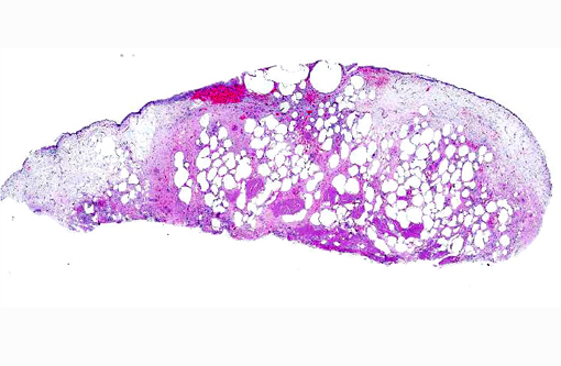
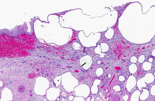
Histopathologic Description:
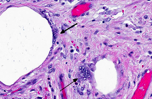
Urinary bladder: The urinary bladder has severe transmural emphysema, severe submucosal edema, mild multifocal submucosal hemorrhage, and a mild, diffuse submucosal inflammation of lymphocytes, plasma cells, eosinophils and macrophages containing hemosiderin. A few macrophages and multinucleate giant cells are present around the air spaces.
Morphologic Diagnosis:
Urinary bladder: Emphysematous cystitis.
Condition:
Emphysematous cystitis
Contributor Comment:
Emphysema of the urinary bladder has been reported in humans, dogs, cattle and cats.(1,2) It occurs with infection of the bladder by gas-producing bacteria that ferment glucose in the urine.
E. coli, Pseudomonas, Klebsiella, Proteus, Enterobacter sp., and
Clostridium sp. are bacteria that have been isolated from cases of emphysematous cystitis in dogs and humans.(1,2) Glucosuria caused by diabetes mellitus is the most common cause of urinary bladder emphysema. The condition also occurs in non-diabetic humans and dogs associated with chronic recurrent cystitis, cortisone administration and primary glucosuria.(1,2)
JPC Diagnosis:
Urinary bladder: Emphysema, transmural, multifocal to coalescing, marked, with mild granulomatous and lymphoplasmacytic cystitis, and submucosal hemorrhage and edema.
Conference Comment:
Conference participants explored various mechanisms contributing to glucosuria and emphysematous cystitis, as summarized above by the contributor. Although diabetes mellitus is the most common underlying cause, proximal renal tubular disorders also result in glucosuria, and may induce emphysematous cystitis in several breeds of dog, including Basenjis, Labrador retrievers, Norwegian elkhounds, schnauzers and Shetland sheepdogs.(4) In Basenjis, this renal tubular abnormality, known as canine Fanconi-like syndrome, is hereditary. It is characterized by impaired renal tubular reabsorption of glucose, phosphate, sodium, potassium, uric acid, amino acids and protein.(6) In humans, Fanconi-Bickel syndrome is due to a defect in the SLC2 gene, which results in defects of GLUT2, a member of the renal glucose transporter protein family.(5) It is suspected that there is a similar underlying defect triggering canine Fanconi-like syndrome, but this has not been proven. Acquired Fanconi-like syndrome in dogs has been associated with several drugs and toxins, such as gentamicin and ethylene glycol. Whether congenital or acquired, Fanconi-like syndrome results in glucosuria (with normal blood glucose), phosphaturia, aminoaciduria and proteinuria.(4) Primary (type II) renal tubular acidosis may also occur due to impaired bicarbonate reabsorption in renal proximal convoluted tubules, which leads to secretional, hyperchloremic metabolic acidosis with normal anion gap and alkaline urine.(3)
Primary renal glucosuria (without other reabsorption abnormalities) is an inherited disorder of Norwegian elkhounds.(4) It is caused by defects in SGLT2, from the sodium-glucose cotransporter family of proteins that is encoded by SLC5 genes in people.(5) As with Fanconi-like syndrome, the underlying genetic defect in dogs has not been established. In addition to primary glucosuria and diabetes, treatment with exogenous corticosteroids, and the subsequent antagonism of insulin, can lead to glucosuria (and thus, emphysematous cystitis) as well; once the resorptive capacity of the proximal renal tubule is exceeded, at a blood glucose concentration greater than 180mg/dL in dogs, glucose spills over into the urine.(3) Glucosuria may also occur in cattle treated with intravenous glucose.
Participants also reviewed other pathologic processes resulting in tissue emphysema. Clostridia are probably the most well-known gas producing organisms, as exemplified by
Clostridium chauvoei induced necrohemorrhagic and emphysematous myositis (blackleg). Pulmonary emphysema is a non-specific change due to alveolar rupture in downer cattle or those with dyspnea or increased respiratory effort. Subcutaneous emphysema in any species could be a sequela to tracheal, bronchial or pulmonary trauma. In swine, intestinal emphysema (pneumatosis cystoides intestinalis) is an idiopathic, incidental finding at slaughter. Fetal death with maceration and emphysema is a fairly well described condition in ruminants. So called gas bubble disease in fish is caused by supersaturated levels of dissolved oxygen or nitrogen in the water of the tank. Lesions are secondary to the accumulation of gas bubbles in blood vasculature and tissues. Finally, postmortem gas production is also a familiar occurrence in veterinary pathology; it can be distinguished from antemortem emphysema by the absence of an inflammatory reaction.
References:
1. Matsuo S, Hayashi S, Watanabe, T, et al. Emphysematous cystitis in a chemically-induced diabetic dog. J Toxicol Pathol. 2009;22:289-292.
2. Lobetti RG, Goldin JP. Emphysematous cystitis and bladder trigone diverticulum in a dog. J Sm Anim Pract. 1998;39:144-147.
3. Latimer KS, ed. Duncan & Prasses Veterinary Laboratory Medicine- Clinical Pathology. 5th ed. Ames, IA: Wiley-Blackwell; 2011:163-165,193-194, 263.
4. Maxie MG, Newman SJ. Urinary system. In: Maxie MG, ed. Jubb, Kennedy, and Palmers Pathology of Domestic Animals. 5th ed., vol. 2. St. Louis, MO: Elsevier Limited; 2007: 474.
5. Santer R, Calado J. Familial renal glucosuria and SGLT2: from a mendelian trait to a therapeutic target. Clin J Am Soc Nephrol. 2010;5(1):133-141.
6. Yearly JH, Hancock DD, Mealey KL. Survival time, lifespan, and quality of life in dogs with idiopathic Fanconi syndrome. J Am Vet Med Assoc. 2004;225(3):377-383.