Signalment:
Gross Description:
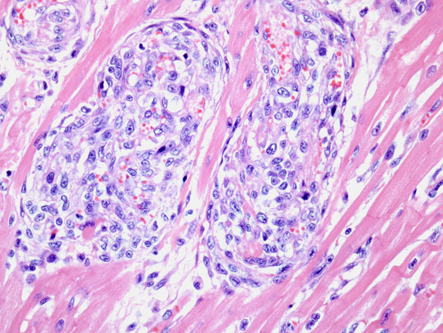
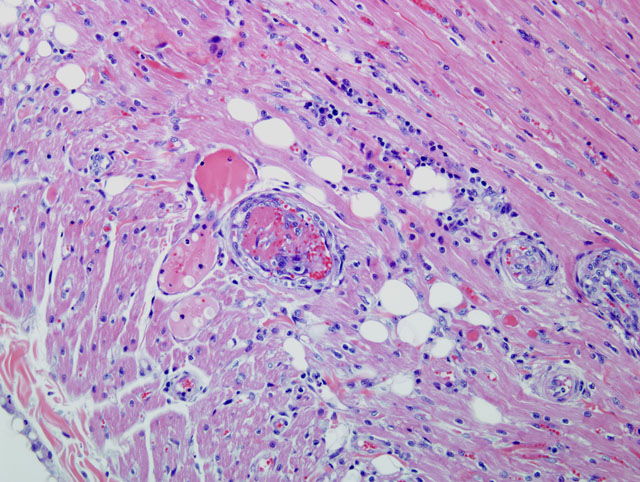
Histopathologic Description:
Similar vascular lesions were present in the kidneys, lungs, pancreas, duodenum, diaphragm, cervical soft tissues, and leptomeninges. Sections from the enlarged spleen and lymph nodes revealed lymphoid hyperplasia.
Immunohistochemistry was performed on heart sections. Most spindle cells in the affected vessels showed membrane-associated expression of factor VIII-related antigen (FVIII-ra, figure 1). Fewer spindle cells showed cytoplasmic expression of smooth muscle actin (SMA, figure 2). The cells did not show expression of CD3 and CD79 (figures 3 and 4).
Morphologic Diagnosis:
Lab Results:
Condition:
Contributor Comment:
Fourteen cases of FSRA have been described, and this appears to be a rare condition affecting exclusively domestic cats.(2,3,4,5) Similar multisystemic vascular lesions have not been described in other animal species. In all reported cases the diagnosis was obtained on post-mortem examination, after the cat died or was euthanized, usually following an acute illness. Affected cats were predominantly young adults, and males appeared more commonly affected. The clinical signs were variable, but most commonly included dyspnea, lethargy and anorexia. Gross lesions were also variable and nonspecific, but included pericardial and pleural effusion, pulmonary edema, and multifocal petechial and ecchymotic hemorrhages of various tissues. An atypical spindle cell proliferation affecting small blood vessels was always observed in the heart. Other commonly affected tissues included kidneys, spleen, lymph nodes, gastrointestinal tract, brain, meninges, eyes, and pancreas. The vascular histologic lesions described in the present case, and the immunohistochemical findings, are similar to those described in the published cases. Ultrastructural examination was described in two cats, and revealed a mixture of two distinct types of spindle cells, consistent with endothelial cells and pericytes.(4,5) The expression of FVIII-ra and SMA is also compatible with a mixed population composed of endothelial cells and pericytes. Based on the presence of two cell types and the lack of cellular atypia, FSRA is believed to represent a reactive proliferative process; it does not appear to be a neoplasm. While the exact cause of death was not clear in most cases, it has been suggested that heart failure probably occurs based on the consistent involvement of the heart, the evidence of perivascular ischemic myocardial necrosis, and the presence of pleural and pericardial effusion, and pulmonary edema with alveolar histiocytosis in many cases.(4)
Histologically and immunohistochemically, FSRA is most similar to reactive angioendotheliomatosis (RAE), a rare human condition.(4) However, RAE in humans is a self-limiting lesion confined to the skin, while FSRA in cats is a multisystemic condition which has been associated with severe illness and death. No similar multisystemic disease has been described in humans. Other human vascular disorders characterized by mixed endothelial cell and pericyte proliferation include intravascular papillary endothelial hyperplasia, acroangiodermatitis (pseudo-Kaposis sarcoma) glomeruloid hemangioma (POEMS syndrome), and some cases of chronic disseminated intravascular coagulation and thrombotic thrombocytopenic purpura.(4) The pathogenesis of these lesions is complex and somewhat distinct for each disease, but possible mechanisms include an exaggerated response to thrombosis, an unusual residuum of immune-mediated vasculitis, and an exuberant angiogenesis possibly related to angiogenic cytokines and a dysfunctional endothelial regulation of coagulation and fibrinolysis.(4) In one case of FSRA, hematologic evaluation showed evidence of thrombotic thrombocytopenic purpura(2), but it remains unclear if this is a significant cause or mechanism in other feline cases. Some proliferative endothelial lesions in human are associated with specific infectious agents, particularly in AIDS patients.(4) Kaposis sarcoma is caused by human herpesvirus-8, and bacillary angiomatosis is caused by Bartonella henselae and B. quintana. In the FSRA cases reported, serologic results were described for only two cats, and there was no evidence of infection by FIV, FeLV, or FIP virus. Silver stains and electron microscopy performed on the lesions from two cats did not show evidence of Bartonella spp. or any other infectious organisms.(4,5) The etiology and pathogenesis of FSRA remains unclear.
The term malignant angioendotheliomatosis has been used to describe intravascular angiotropic lymphoma in humans and animals. In humans, RAE has historically been confused with intravascular lymphoma. This case was not consistent with lymphoma based on cell morphology, and this was confirmed by the lack of expression of CD3 and CD79.
JPC Diagnosis:
Conference Comment:
The pathogenesis of FSRA is still unknown, but it is hypothesized that a reactive response to thrombosis, vasculitits, or an infectious agent is the cause.(1)
References:
2. Cooley AJ, Rushton SD, Porterfield ML, Tice CA: Arteriolar endothelial proliferation and microthrombosis attributed to thrombotic thrombocytopenic purpura in two cats. Vet Pathol 41: 576, 2004
3. Dunn KA, Smith KC, Blunden AS: Fatal multisystemic intravascular lesions in a cat. Vet Rec 140:128- 129, 1997
4. Fuji RN, Patton KM, Steinbach TJ, Schulman FY, Bradley GA, Brown TT, Wilson EA, Summers BA: Feline systemic reactive angioendotheliomatosis: Eight cases and literature review. Vet Pathol 42: 608-617, 2005
5. Rothwell TL, Xu FN, Wills EJ, Middleton DJ, Bow JL, Smith JS, Davies JS: Unusual multisystemic vascular lesions in a cat. Vet Pathol 22:510-512, 1985