Signalment:
Gross Description:
Histopathologic Description:
Morphologic Diagnosis:
Lab Results:
Condition:
Contributor Comment:
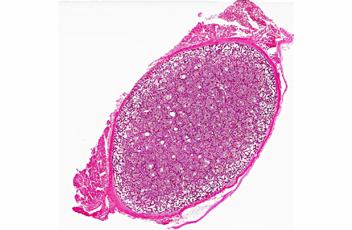
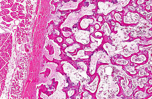
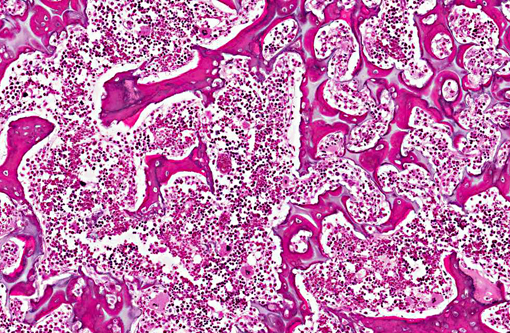
Gross diagnosis is established by splitting long bones and demonstrating persisting primary spongiosa in the medullary cavity. Histologically, osteoclasts are markedly reduced in number, in spite of the presence of resorption lacunae. In this section of rib there are scant osteoclasts. Where present, they are lightly stained with washed out vesicular nuclei.
The basis for the defect is a mutation in the anion exchanger protein coded for by SLC4A2 on chromosome 4. A similar mutation causes osteopetrosis in mice.(1,7) To date, no comparable mutation has been recognized in severe forms of osteopetrosis in children. Human osteopetrosis is classified morphologically into forms with reduced, normal or excessive numbers of osteoclasts. In Angus cattle it is likely that absence of the anion exchanger results in alkalinized cytoplasm, resulting in death of osteoclasts.(3) Death in utero is due to compression of the cerebral cortex with posterior herniation of cerebellum as a result of osseous crowding in the cranial vault.(4) An important diagnostic rule out is osteopetrosis due to in utero infection with bovine viral diarrhea virus. In this instance this and other calves sired by the bull were negative for BVDV.
JPC Diagnosis:
Conference Comment:
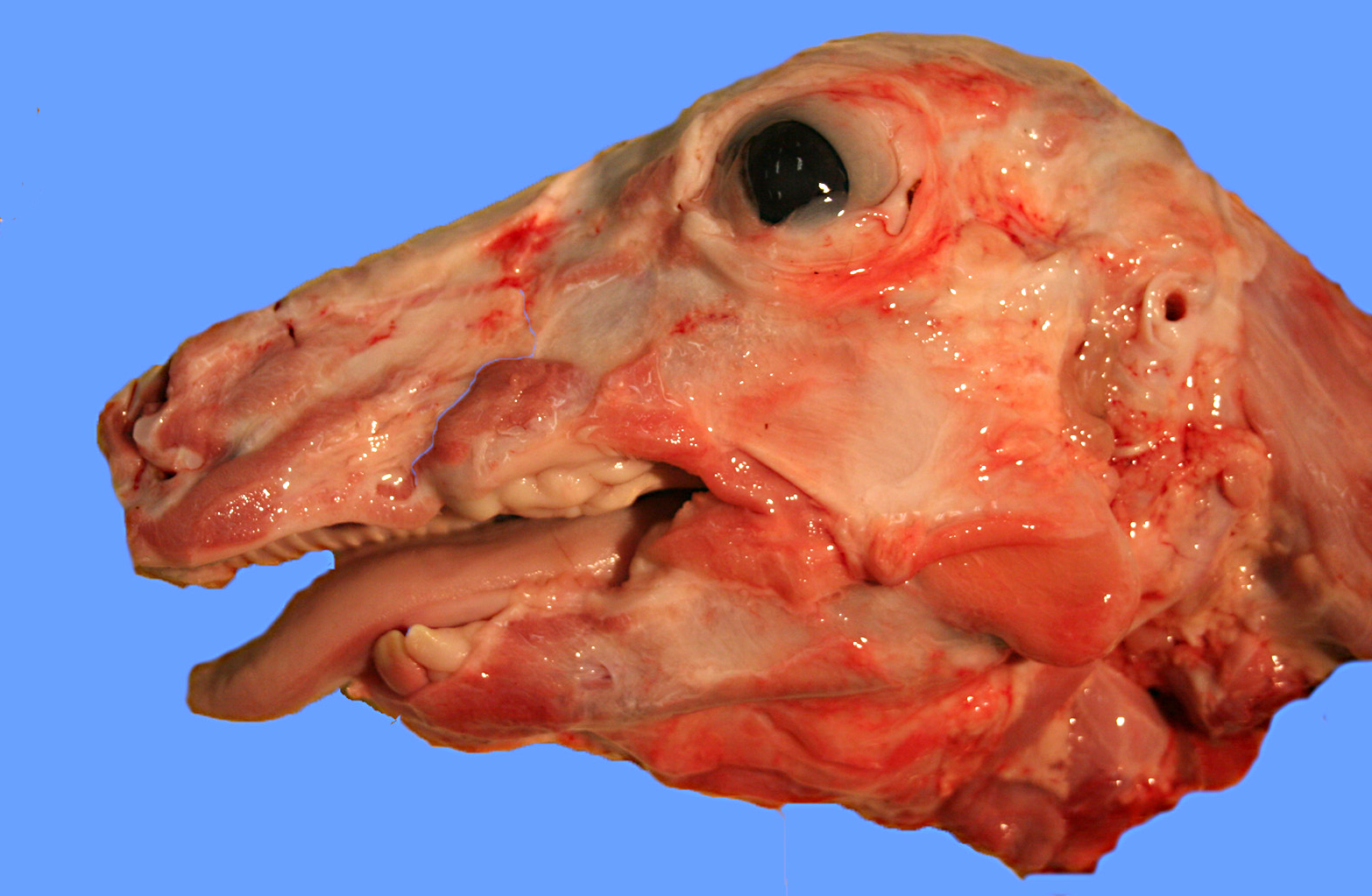
The distinctive gross appearance of affected calves identified by the contributor are:
- compression of cerebral hemispheres with herniation of the cerebellar vermis into the foramen magnum
- dorsoventrally compressed brain with a concave depression in the parietal cortex
- thickened parietal bone
- mildly proptosis
- protruding tongue
- brachygnathia inferior
- restricted movement of temporomandibular joints
- loose, misaligned, not fully erupted inscisors
- misaligned and unerupted molars
- abnormally smooth cranial calvarium surface with prominent bilaterally symmetrical bony ridges at the frontal-parietal synchondrosis and a corresponding depression in the temporoparietal cortex
- 1-2 cm fontanelle between frontal and parietal bones
- bilaterally symmetrical transverse ledge of bone at the frontalsphenoid synchondrosis that involved the orbital wing of the sphenoid bone4
Distinctive histologic findings are:
- trabecular bone with cores of hyaline cartilage
- rare osteoclasts in long bones, with numerous osteoclasts in the skull
- significant reduction or absence of retinal ganglion cells
- axonal loss and gliosis in the optic nerves and optic chiasm
- chromatolysis and neuronal atrophy in nuclei of the medulla oblongata (facial, vestibulocochlear, and hypoglossal nuclei, and red nuclei)
- atrophy of muscles of the tongue and larynx
- axonal swelling and Wallerian degeneration in pyramidal tracts and medial longitudinal fasciculus perivascular paraventricular corpora amylacea in the thalamus, basal nuclei, and midbrain with similar but larger structures in the choroid plexus of the third ventricle
- calcified vessel walls and mineralization of neuropil associated with the corpora amylacea
- reduced Purkinje cell numbers (but no other histologically identifiable cortical atrophy in compressed parietal cortex or in herniated cerebellum)
- disorganized mixture of cementum, dentin, enamel and trabecular bone in teeth (particularly around apical foramina within the intra-alveolar portion of premolars)
- mineralized and ossified connective stroma in the tooth chamber
- periodontal hemorrhage
- increased ratio of acellular to cellular cementum4
Conference participants noted that the cortex in the examined sections appears thin. Since we are unclear as to the level at which these sections were taken, we cannot fully evaluate this cortical thinning. It was speculated that the sections may be through the cutback zone, in which case the cortex would be expected to be discontinuous and thin.
References:
2. Leipold HW, Huston K, Dennis SM, Guffy MM. Hereditary osteopetrosis in Aberdeen Angus Calves. I. Pathological changes. Ann G+â-¬n+â-¬t S+â-¬l Anim. 1971;3:245-253.
3. Meyers SN, McDaneld TG, Swist SL, et al. A deletion mutation in bovine SLC4A2 is associated with osteopetrosis in Red Angus cattle. BMC Genomics. 2010;11:337.
4. OToole D, Swist S, Steadman L, Johnson GC. Neuropathology and craniofacial lesions of osteopetrotic Red Angus calves. Vet Pathol. 2012;49:746.
5. Thompson K. Diseases of bones and joints. In: Maxie MG, ed. Pathology of Domestic Animals. 5th ed. Vol. 1. Edinburgh, Scotland: WB Saunders; 2007:38-40.
6. Thomson RG. Failure of bone resorption in a calf. Vet Pathol. 1966;3:234-246.
7. Wu J, Glimcher LH, Aliprantis AO. HCO3-/Cl- anion exchanger SLC4A2 is required for proper osteoclast differentiation and function. Proc Natl Acad Sci USA. 2008;105:16934-16939.