Signalment:
Gross Description:
Histopathologic Description:
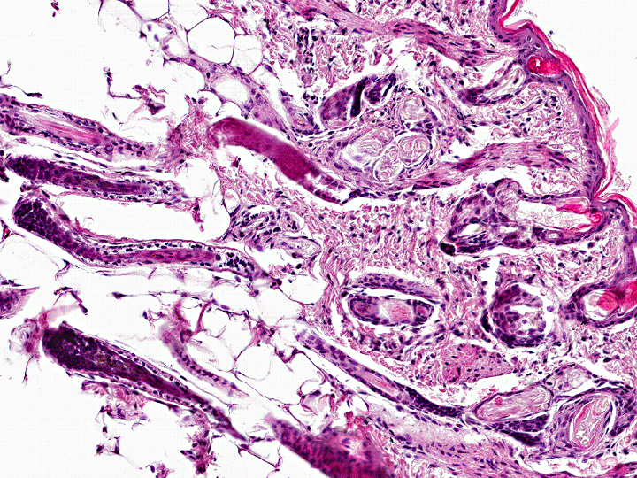
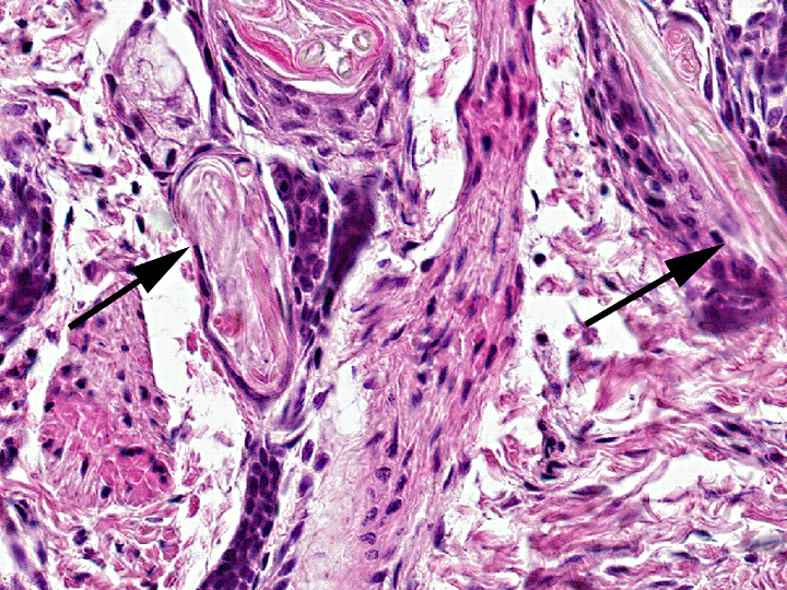
Few mild lymphoplasmacytic infiltrates are present in the superficial and perifollicular dermis. The mostly bilayered epidermis is unremarkable with multifocal to segmental mild orthokeratotic hyperkeratosis.
Morphologic Diagnosis:
Condition:
Contributor Comment:
The main clinical feature, alopecia occurring at or shortly after birth, can be induced by changes in either quantity or quality of hair shafts. Congenital changes are based most frequently on inherited abnormal morphogenesis and therefore are termed hair follicle dysplasias. There are several kinds of dysplasias, depending to the differentiation level of affected cells (single differentiated cell or progenitor cell). Accordingly, changes in related tissues like cutaneous appendages, nails or claws and teeth may occur.
The hair follicle with its product, the hair shaft, is a complex unit underlying tightly regulated cyclic changes.(6) Classification relies on differentiation between reduced hair follicle quantity and quality.(7)
Alterations in hair follicle quantity: Developmental reduction of hair placodes during organogenesis or defective morphogenesis with permanent loss of hair follicles can both lead to congenital alopecia. Delineation is often difficult because variable subsets of hair follicles are existent and are irregularly distributed over different body regions.
Aplasia of hair follicles with dental dysplasia affects more than one tissue derived from the ectoderm, mostly apocrine glands. Several forms have been reported in humans, various breeds of dogs,(10) mice,(2) and cattle,(1) with only few of the underlying signaling pathway defects characterized yet.(2) Aplasia of hair follicles without dental dysplasia with a dominant autosomal inheritance has been described in pigs.(7)
Alterations in hair follicle quality: Structural changes leading to reduced, defective or absent hair shaft production can be subdivided into those resulting from morphological changes of the hair follicle itself or those where morphologically unchanged hair bulbs form defective hair shafts.
Especially in the dog, several breeds are affected with hairlessness resulting from hair follicle dysplasia with defects in hair follicle development, for example Mexican(5) or Peruvian hairless dogs and Chinese crested dogs.(11) In contrast to horses with only rare reports, diverse forms have been described in various breeds of cattle. Some of these have already been characterized as autosomal recessive or dominant, partly lethal traits.(7)
Hair follicle dysplasia without defects in hair follicle development (trichomalacia) is classically represented by nude mice amongst few others.(8) Beside few breeds of cats, (e.g. Sphinx breed) and dogs,(7) mainly in man, numerous forms of trichomalacia have been observed.(4)
A special form of dysplasia affects the neuroectodermally derived follicular melanocytes which also contribute to regular hair follicle development.(3) Histopathological changes are specific and identical in both syndromes with formation of enlarged melanin granules in melanocytes and later aggregation of perifollicular melanophages.(7) Depending on the affected breed and coat colors, color dilution alopecia and black hair follicular dysplasia have been differentiated.(13)
The case submitted here fulfills the relevant criteria for hair follicle dysplasia without defects in hair follicle development (trichomalacia) because of the unchanged appearance of hair follicles. Further changes in ectodermderived tissues, like apocrine glands or teeth, were not observed. The occurrence in both animals of the litter is suggestive of an inherited genetic defect. The queen had been mated to the identical sire before, without any abnormality in the offspring. Unfortunately the littermate was not available for pathological examination and confirmation of similar changes in hair shaft formation.
Death of this kitten was attributed to acute hepatic failure with severe hepatocellular degeneration and lipidosis (peripheral lipomobilization syndrome) based on the results of pathological examination of all other organ systems, including histology.
JPC Diagnosis:
Conference Comment:
References:
2. Freire-Maia N, Lisboa-Costa T, Pagnan NAB: Ectodermal dysplasias: How many? American Journal of Medical Genetics 104: 84-84, 2001
3. Hume AN, Tarafder AK, Ramalho JS, Sviderskaya EV, Seabra MC: A Coiled-Coil Domain of Melanophilin Is Essential for Myosin Va Recruitment and Melanosome Transport in Melanocytes. Mol Biol Cell 17: 4720-4735, 2006
4. Itin PH, Fistarol SK: Hair shaft abnormalities--clues to diagnosis and treatment. Dermatology 211: 63-71, 2005
5. Kimura T, Ohshima S, Doi K: The Inheritance and Breeding Results of Hairless Descendants of Mexican-Hairless Dogs. Laboratory Animals 27: 55-58, 1993
6. Krause K, Foitzik K: Biology of the hair follicle: The basics. Seminars in Cutaneous Medicine and Surgery 25: 2-10, 2006
7. Mecklenburg L: An overview on congenital alopecia in domestic animals. Vet Dermatol 17: 393-410, 2006
8. Mecklenburg L, Tychsen B, Paus R: Learning from nudity: lessons from the nude phenotype. Experimental Dermatology 14: 797-810, 2005
9. Meckelburg L. Trichomalacia. In: Meckelburg L, Linek M, Tobin DJ, eds. Hair Loss Disorders in Domestic Animals. Ames, IA: Wiley-Blackwell; 2009:115-117.
10. Moura E, Cirio SM: Clinical and genetic aspects of X-linked ectodermal dysplasia in the dog -a review including three new spontaneous cases. Veterinary Dermatology 15: 269-277, 2004
11. Sander P, Drogemuller C, Cadieu E, Andre C, Leeb T: Analysis of the canine EDAR gene and exclusion as a candidate for the hairless phenotype in the Chinese Crested dog. Animal Genetics 36: 168-171, 2005
12. Tieghi C, Miller WH Jr, Scott DW, Pasquinelli G. Medullary trichomalacia in 6 German shepherd dogs. Can Vet J. 2003 Feb; 44(2):132-6.
13. Welle M, Philipp U, Rufenacht S, Roosje P, Scharfenstein M, Schutz E, Brenig B, Linek M, Mecklenburg L, Grest P, Drogemuller M, Haase B, Leeb T, Drogemuller C: MLPH Genotype--Melanin Phenotype Correlation in Dilute Dogs. J Here'd, 2009