CASE I: 288/09 (JPC 41120035-00)
Signalment:
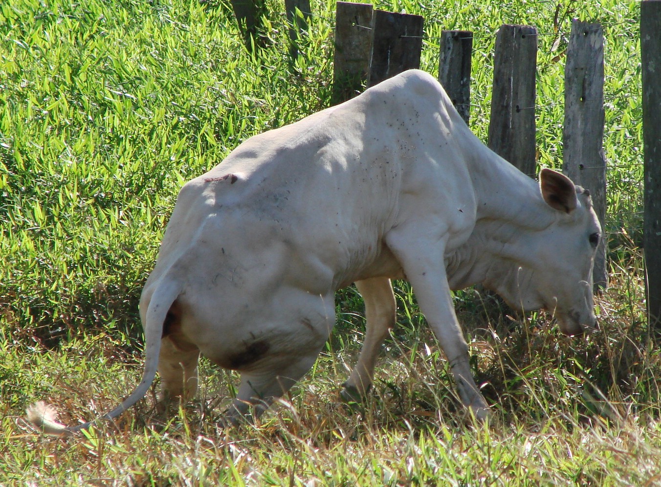
24-month old, male, Nelore (Bos taurus indicus), bovine.
History:
The disease occurred in January-February 2010, in cattle held on a farm in the state of Pará Norh Brazil. Twenty five out of 3,000 cattle were affected by a neurological disorder 60 days after being vaccinated against the foot and mouth disease (FMD). The affected cattle were of different ages and fair nutritional condition. They presented a 5cm diameter nodular protuberance, commonly on the left side of the lumbar region midline, involving muscle and subcutaneous tissue. The clinical signs were consistent with lumbar spinal cord compression syndrome with progressive paralysis of the hind limbs, characterized by ataxia with signs of dragging the hooves, crossing of hindlimbs when walking, "dog sitting" position with spread hindlimbs, urinary incontinence, and stumbling and falling. These signs lasted for 2-5 months and evolved to sternal recumbency with difficulty in rising. The sensory and motor reflexes of the forelimbs were normal.
Gross Pathology:
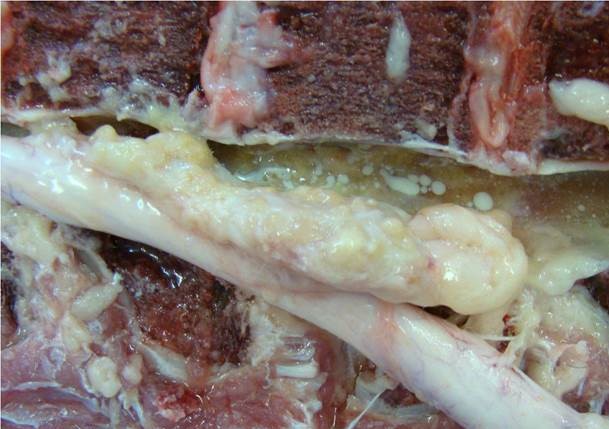
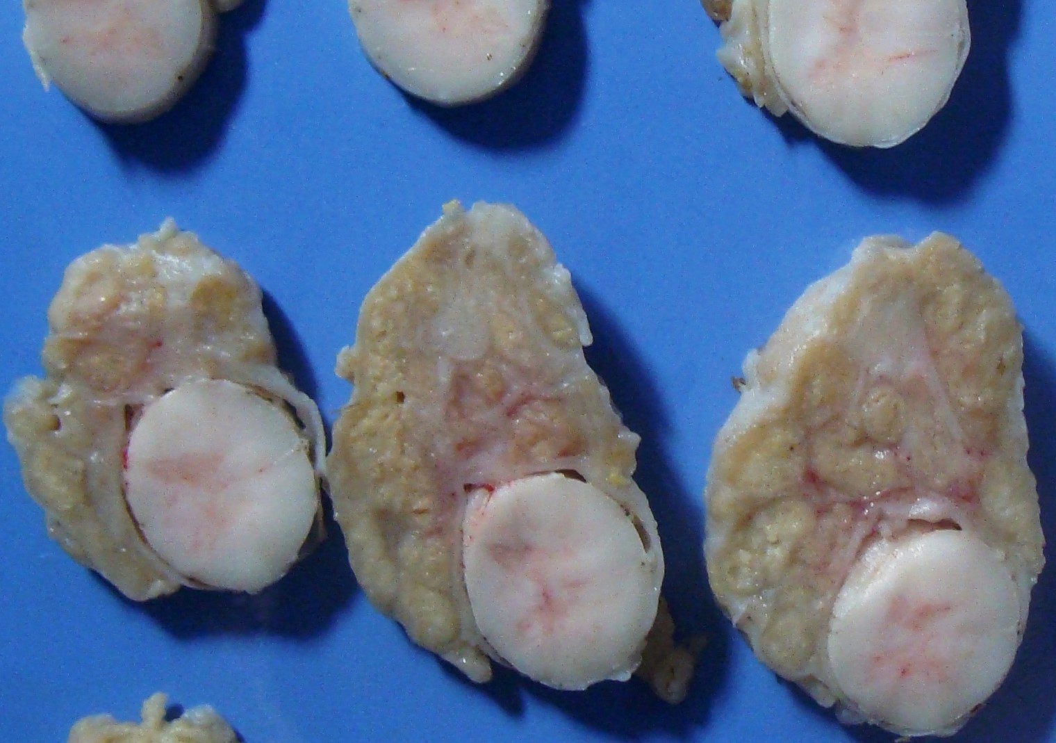
There were areas of grayish yellow discoloration in the longissimus dorsi muscle. These areas were firm and infiltrated muscle bundles and communicated with a roughly fusiform cylindrical 10x3cm white-yellowish mass located in the epidural space extending from L1 to L4 and adhered both to the periosteum of the vertebral body and to the dura mater (Fig. 2). In this area, whitish oily fluid, resembling the consistency of FMD vaccine, was infiltrated the intervertebral spaces. On cut surface (Fig. 3), the epidural mass was white and firm and had multifocal to coalescing, soft, fairly nodular yellow foci ranging in size from 0.2 to 2 cm in diameter.
Laboratory Results:
A myelography of the lumbosacral revealed contrast retention at the level of L4 vertebra and an intradural mass extending from L1 to L3. No bacterial growth was obtained from the bacterial culture of spinal cord samples from this region.
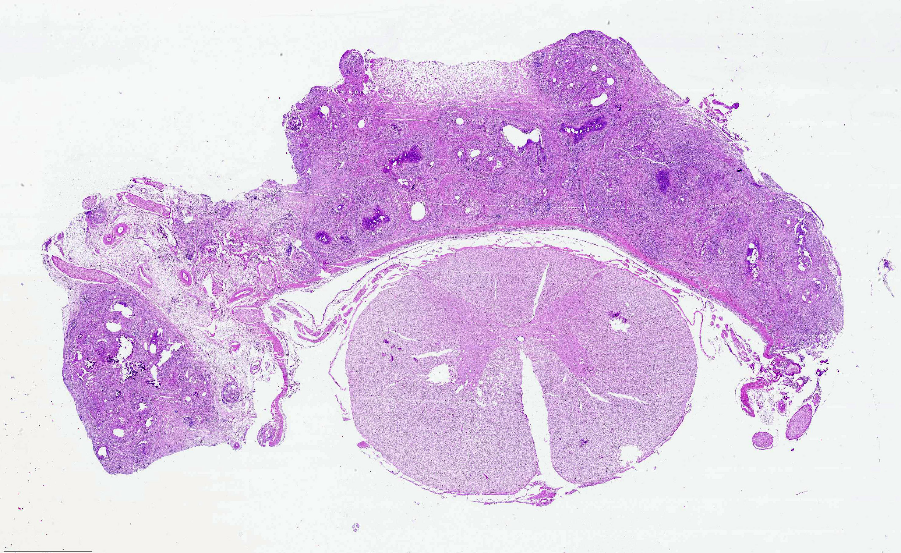
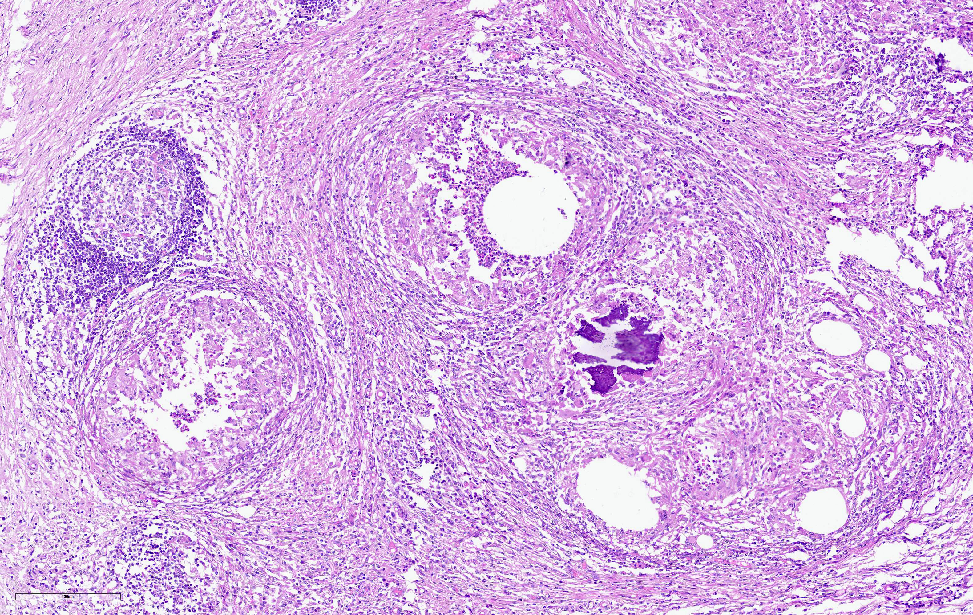
Microscopic Description:
The subcutaneous tissue of the lumbar area and muscle was expanded and partially effaced by extensive pyogranulomatous inflammation and fibrosis. There was hyaline and floccular degeneration of the striated muscle in the area. The mass in the epidural space consisted of the same type of pyogranulomatous inflammation. Some of the pyogranulomas consisted of a necrotic and mineralized with numerous neutrophils surrounded by foamy and epithelioid macrophages (occasionally forming multinucleated giant cells), lymphocytes, and plasma cells. Clear spaces ranging from 20 to 200μm were seen in central necrotic areas. The dura mater was thickened by fibrosis and the epidural space obliterated the pyogranulomatous mass. Neuronal chromatolysis were observed mainly in the dorsal horns of the gray matter of the spinal cord, and mild to severe Wallerian degeneration in the dorsal funiculi.
Samples from the granulomatous mass and spinal cord were stained using Steiner's silver and Ziehl-Neelsen acid-fast techniques.
Contributor's Morphologic Diagnosis:
1. Severe, focally extensive, pyogranulomatous panniculitis; myositis with intralesional vacuoles.
2. Spinal cord pyogranulomatous pachymeningitis with intralesional vacuoles (consistent with oil adjuvant droplets).
3. Myelomalacia, secondary to spinal cord compression (consistent with oil adjuvant droplets).
Contributor's Comment:
The findings described here characterize cases of spinal cord compression caused by granulomatous inflammation caused by incorrect inoculation of the FMD vaccine. Vaccines against FMD contain an oil adjuvant. Adjuvants act non-specifically by increasing the immune response against antigens and are a key component of vaccines. They can cause adverse effects such as anaphylaxis, infection, granulomas at the injection site, and tumors.4
The cases of spinal cord compression described here were attributed to improper vaccination. There is limited information about economic losses due to vaccine reactions in cattle in Brazil. The practice of mass vaccination, mainly in beef cattle, has led to the appearance of abscesses in the injection site because of improper sharing of needles between individuals and antisepsis procedures during the vaccination.
The most remarkable histopathological findings in this case were somewhat similar to those observed in others studies.3,5,6,7 Demonstration of lipid-like material surrounding or within the center of pyogranulomas, coupled with the absence of bacterial infection, indicated that the reaction was most likely directed toward a component in the vaccine, in association with the practice of improper procedures.
In domestic animals, which exhibit posterior ataxia and paralysis, several differential diagnoses such as fractures, neoplasms (mainly lymphosarcoma), rabies, and botulism should be considered in this region of Brazil. However, most of them have clinical, macroscopic and microscopic different aspects, which were ruled out. In this case, the history of incorrect vaccination procedures was essential to the diagnosis.7
The neck is the usual inoculation site for the FMD vaccine in cattle in Brazil. Paravertebral intramuscular injection sites should be avoided for vaccination purposes when potentially irritating biological products are used.
Contributing Institution:
Laboratório de Patologia Veterinária, Universidade Federal de Mato Grosso
http://www1.ufmt.br/ufmt/unidade/?l=ppgvet
JPC Diagnosis:
Spinal cord, meninges and epidural fat: Pyogranulomas, multiple, with numerous clear vacuoles (consistent with oil droplets) and marked fibrosis.
JPC Comment:
The contributor's comment provides a concise overview of adjuvant's role within vaccines and potential consequences of inappropriate administration.
Adjuvants (Latin: adjuvare "to help") mimic microbial antigens detected by antigen presenting cell pattern recognition receptors, increasing the innate immune system's response and subsequently facilitating the development of adaptive immunity. Antigen presenting cells subsequently express of molecules called costimulators and secrete cytokines that stimulate the proliferation and differentiation of T lymphocytes in peripheral lymphoid organs which then facilitate development of the adaptive immune system (i.e. cell mediated and humoral immunity).2,5
Water-in-oil adjuvanted vaccines are commonly used in food animals worldwide, including the United States, but are no longer used in human formulations due to their irritancy in tissue, particularly in regard to more slowly absorbed mineral oil mixtures.5
Scar tissue formation is an undesirable side effect of vaccination site reactions in food animals, reported to cost the United States cattle industry $55 million dollars annually (2005 value) due to trim, affecting 10% of carcasses in 1998.5 Producer organizations in North America therefore recommend biological, antibiotic, vitamin, and mineral products be formulated for subcutaneous use whenever possible and administered in the neck. However, these products may ultimately be administered elsewhere, such as in epaxial or hip muscles for reasons such as operator safety, ease of administration, and in some cases lack of a manufacturer statement advising against injection in these locations.5
Aqueous based (often combined aluminum hydroxide [Al(OH)3] and saponin) adjuvants are also commonly used in veterinary medicine and have also been implicated in the formation of immune mediated granulomas.1,8
One study in sheep found approximately 92% of lambs injected with an aluminum based adjuvant without vaccine solution developed immune mediated granulomas. The presence of aluminum within granulomas (and lymph nodes) was confirmed using fluorescence microscopy with lumogallion staining and electron microscopy.1
The moderator discussed additional causes of compressive myelopathy in ruminants, including abscesses, trauma, malformations, and neoplasia.
References:
1. Asín J, Molín J, Pérez M, et al. Granulomas Following Subcutaneous Injection With Aluminum Adjuvant-Containing Products in Sheep. Vet Pathol. 2019;56(3):418-428.
2. Diseases of the immune system. In: Kumar V, Abbas AK, Aster JC, eds. Robbins and Cotran Pathologic Basis of Disease. 9th ed. Philadelphia, PA: Elsevier; 2015:189-204.
3. Kleinman NR, Kier AB, Diaconu E, et al. Posterior paresis induced by Freund's adjuvant in guinea pigs. Lab Anim Sci. 1993;43(4):364-366
4. O'Toole D, McAllister MM, Griggs K. Iatrogenic compressive lumbar myelopathy and radiculopathy in adult cattle following injection of an adjuvanted bacterin into loin muscle: Histopathology and ultrastructure. J. Vet Diagn Invest 1995;7(2):237-244
5. O'Toole D, Steadman L, Raisbeck M, et al. Myositis, lameness, and recumbency after use of water-in-oil adjuvanted vaccines in near-term beef cattle. J Vet Diagn Invest. 2005; 17(1):23-31
6. Panziera W, Rissi DR, Galiza GJN, et al. Pathology in practice. J Vet Med Assoc. 2016; 249 (5): 483-485.
7. Ubiali DG, Cruz RAS, Lana MVC, et al. Spinal cord compression in cattle after the use of an oily vaccine. Pesq Vet Bras. 2011; 31(11): 997-999.
8. Ulziibat G, Maygmarsuren O, Khishgee B, et al. Immunogenicity of imported foot-and-mouth vaccines in different species in Mongolia. Vaccine. 2020;38(7):1708-1714.