Signalment:  

Ten-week-old, female, border collie dog Canis familiaris.The 10 week old puppy was euthanized and submitted for necropsy with a history of ascending progressive muscle weakness, neutrophilic leukocytosis, non-regenerative anemia, and elevated alkaline phosphatase levels.


Gross Description:  

The carcass was in good physical condition and post mortem autolysis was mild. Mucopurulent ocular discharge and crusts were present bilaterally. Mucus membranes, subcutaneous tissues and viscera were uniformly pale. Small numbers of petechia were present in the peritoneum overlying the ventral abdominal muscles. A diffuse copper tint was present throughout the liver parenchyma. Intestinal contents were bright yellow and watery. No additional significant gross abnormalities were identified. 


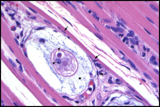
Histopathologic Description:

Skeletal muscle: Endomysial areas are infiltrated by scattered small mixed populations of neutrophils and macrophages. Combinations of macrophages and neutrophils also commonly form multifocal, large, vascularized nodular aggregates containing myofiber fragments. Within these pyogranulomas, macrophages often possess an eccentric nucleus due to the presence of single, small, round, amphophilic, intracytoplasmic parasite. Scattered throughout are numerous, round to oval, 50-100 _m structures containing a central, finely granular, pale eosinophilic core, with 1-2 ill-defined nuclei. Surrounding the core are multiple layers of lacy, pale basophilic material and a thin outer wall of flattened cells with elongated nuclei (onion cysts). These structures elicit little or no direct inflammatory response. Present in some sections are similarly sized structures (meronts) with a large pale eosinophilic core lined peripherally by either marginated nuclear material or elongated bodies (merozoites) with deep basophilic nuclei. Blood vessels contain increased numbers of neutrophils and macrophages.


Morphologic Diagnosis:  

Skeletal muscle: Myositis, pyogranulomatous, widespread, chronic, severe, with multifocal intralesional H. americanum zoites and meronts


Condition:  

Hepatozoon americanum


Contributor Comment:  

The history and histopathologic findings are consistent with American canine hepatozoonosis, a debilitating, tick-borne disease of dogs in the south-central and southeastern United States. The diagnosis was confirmed by PCR and sequence analysis. Discovered in 1978, H. americanum was advanced as a distinct species from its Old World counterpart, H. canis, in 19971,8. At least 46 Hepatozoon species infect mammals and more than 120 infect snakes. Transmission of the apicomplexans to their vertebrate intermediate hosts occurs through hematophagous invertebrates. Like their Plasmodium and Babesia spp. relatives, many hepatozoons occur in erythrocytes. However, most of the mammalian parasites infect leukocytes and use acarines as definitive hosts or vectors. Interestingly, H. americanum is spread by the ingestion of infected ticks rather than through their feeding activities3.

It is believed that H. americanum crossed into canids from unknown vertebrates only recently, whereas H. canis has a long history with dogs. The vector for H. americanum is Amblyomma maculatum, gamonts are found in monocytes, and merogony occurs in host cells lodged between striated muscle fibers. In contrast, Rhipicephalus sanguineus is a primary vector for H. canis, the neutrophil is the favored host cell, and merogony takes place in a wide variety of tissues. Meronts of H. americanum develop most consistently in striated muscle within onion skin cysts created by layers of host secreted mucopolysaccharide. No similar lesion is associated with H. canis, which rarely occurs in muscle. Disease from H. americanum is more severe than that seen with H. canis. Developing organisms are shielded from the dogs immune system, but elicit intense local pyogranulomatous inflammation, systemic reaction, and overt illness when merozoites are released. Local lesions evolve into vascular granulomas. Diseased dogs are often febrile and lethargic, with stiff gaits, mucopurulent ocular discharge, and atrophy of the head muscles. Clinicopathological findings include mature neutrophilia, increased alkaline phosphatase, and hypoalbuminemia. Periosteal bone proliferation may be seen radiographically and at necropsy2,3,5. 


JPC Diagnosis:  

Skeletal muscle: Myositis, pyogranulomatous, multifocal, moderate, with fibrosis, and intracellular protozoal cysts and zoites etiology consistent with Hepatozoon americanum, Border collie (Canis familiaris), canine.


Conference Comment:  

The disease course of Hepatozoon canis, the causative agent of Old World hepatozoonosis, is generally mild with low levels of parasitemia, but severe illness occurs occasionally with nearly 100% of neutrophils containing parasites.1,2 In the case of Hepatozoon americanum infection, the causative agent of American canine hepatozoonosis, parasitemia remains very low, and often less than 0.1% of leukocytes are infected, even in cases of severe illness.1,2

When H. americanum was first identified, Rhipicephalus sanguineus was thought to be the vector for transmission, as it is a known vector for H. canis. Even current literature has implied the role of R. sanguineus in transmission of H. americanum.6 In fact, the primary vector for H. americanum appears to be Amblyomma maculatum, as R. sanguineus, Dermacentor variabilis, and Amblyomma americanum appear refractory to infection.3,8

In addition to the lesions within skeletal and cardiac muscle, H. americanum is known to cause severe periosteal bone proliferation of proximal limbs.3 Flat bones can be markedly, but less commonly, affected, and distal limbs are often spared.3 The periosteal reaction shares common features with hypertrophic osteopathy in dogs.3 The pathogenesis of the reaction is unknown, as there are no parasites identified with the bone lesions, and the inciting factors have not been identified. 

Severe muscle wasting, especially of the temporal muscles, is also a feature of H. americanum.3

We thank Dr. C. H. Gardiner, PhD, veterinary parasitology consultant to the AFIP, for his review of this case.


References:

1. Baneth G, Barta JR, Shkap V, Martin DS, MacIntire DK, and Vincent-Johnson N: Genetic and antigenic evidence supports the separation of Hepatozoon canis and Hepatozoon americanum at the species level. J Clin Microbiol 38:1298-1301, 2000.
2. Baneth G, Mathew JS, Shkap V, MacIntire DK, Barta JR, and Ewing SA: Canine hepatozoonosis: two disease syndromes caused by separate Hepatozoon spp. Trends in Parasitology 19:27-31, 2003.
3. Ewing SA and Panciera RJ: American canine hepatozoonosis. Clin Microbiol Rev 16:688-697, 2003.
4. Gardiner CH, Fayer R, Dubey JP: An Atlas of Protozoan Parasites in Animal Tissues, 2nd ed., p. 4. Armed Forces Institute of Pathology, Washington, DC, 1998
5. Panciera RJ, Mathew JS, Cummings CA, Duffy JC, Ewing SA, and Kocan AA: Comparison of tissue stages of Hepatozoon americanum in the dog using immunohistochemical and routine histologic methods. Vet Pathol 38:422-426, 2001.
6. Valentine BA, McGavin MD: Skeletal muscle. In: Pathologic Basis of Veterinary Disease, eds. McGavin MD, Zachary JF, 4th ed., pp. 1035-1037. Elsevier, St. Louis, MO, 2007
7. Van Vleet JF, Valentine BA: Muscle and tendon. In: Jubb, Kennedy, and Palmers Pathology of Domestic Animals, ed. Maxie MG, 5th ed., vol. 1, pp. 270-271. Elsevier Limited, St. Louis, MO, 2007 8. Vincent-Johnson NA, MacIntire DK, Lindsay DS, Lenz SD, Baneth G, Shkap V, and Blagburn BL: A new hepatozoon species from dogs: description of the causative agent of canine hepatozoonosis in North America. Am J Parasitol 83:1165-1172, 1997. 

A virtual slide is not available for this case.


Skeletal muscle, Boxer


Back | VP Home | Contact Us |