Signalment:
6-week-old female mixed breed horseFive day history of lameness of right rear leg. Radiographic findings were interpreted to represent septic arthritis of the right coxofemoral joint.
Gross Description:
The foal was in good post mortem condition and had a body condition score of 3/5. Approximately 50% of the cartilage of the right femoral head was ulcerated. On cross section, the subchondral bone underlying the ulcerated articular cartilage was white for a distance of up to 2 mm from the surface. The dorsolateral aspect of the right femoral head had a large cartilage flap that was attached at the margins of the articular surface, and the ventrolateral aspect of the right femoral head had a 4 cm long fissure within articular cartilage that remained attached to subchondral bone. The dorsal region of the acetabular cartilage was irregular with exposure of subchondral bone (ulceration). The right medial trochlear ridge had 2 cm focus of articular cartilage that extended 1 cm into subchondral bone (found on cutting in a horizontal plane using a band saw). The contralateral coxofemoral joint, stifle joint, and both humeral joints were examined and found to be within normal limits.Â
Histopathologic Description:
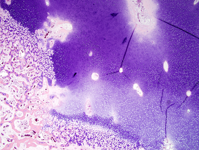
Medial trochlear ridge of the right femur: There is a rectangular area of epiphyseal growth cartilage (AE complex) which extends deeper into the epiphysis (retained cartilage) compared with the adjacent cartilage(
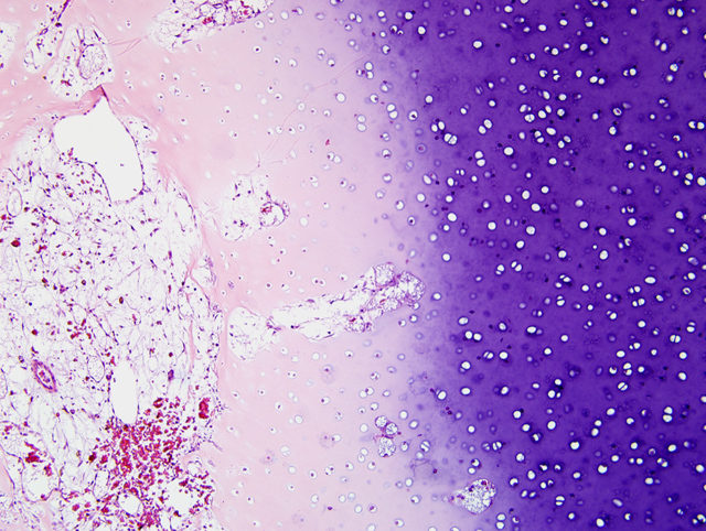
Fig. 1-1). The deep edge of this retained cartilage has cartilage cores present indicating active cartilage mineralization and vascular invasion as seen in endochondral ossification. The chondrocytes in this retained cartilage and the chondrocytes contiguous within its overlying growth cartilage have eosinophilic chondrocytes interpreted as chondrocyte coagulation necrosis compared with chondrocytes not associated with the retained region. In the overlying epiphyseal cartilage, in addition to chondrocyte coagulation necrosis, is a focus of loss of basophilia of the cartilage matrix (loss of proteoglycan staining). The blood vessels in the cartilage canals within this region of proteoglycan loss appear normal, but the contents of some of the cartilage canals in the contiguous growth cartilage with dead chondrocytes appear necrotic compared with contents of cartilage canals in adjacent cartilage with viable chondrocytes(
Fig. 1-2). The primary trabeculae deep to the area of retained cartilage are course and occasionally fractured. In most of these trabeculae the bone is dead (karyolytic osteocytes) often with overlying viable woven bone. There is mild to moderate reactive fibrosis in the marrow in this region.
Condition:
Osteochondrosis
Contributor Comment:
This lesion represents articular epiphyseal complex dysplasia of osteochondrosis. This lesion likely was not clinically significant. The clinical signs in this case were attributed to the lesions in the femoral head which, after microscopic examination, were interpreted to be sequelae to suppurative osteomyelitis. The lesions in the distal femur were found on gross examination of sawed sections to check for foci of osteochondrosis.Â
Using terminology suggested in recent literature, the lesion in the distal femur is a good example of chondrocyte coagulation necrosis in both a focus of osteochondrosis latens and osteochondrosis manifesta.(3) Often the chondrocyte coagulation necrosis is limited to the focus of osteochondrosis latens and it is hypothesized that this necrosis is secondary to ischemia and might be the initiating lesion of osteochondrosis. In the foal as in other species, it has been suggested that this cartilage necrosis is secondary to necrosis of blood vessels in the cartilage canals of growth cartilage.(1) Interesting in this lesion is the presence of cartilage cores at the deep margin of the retained cartilage. This indicates cartilage mineralization and vascular invasion are taking place. This is not usually found in lesions of osteochondrosis manifesta. Osteochondrosis manifesta lesions are known to be able to resolve (2); therefore active endochondral ossification might represent attempts to resolve the lesion. The abnormal modeling, fractures and marrow fibrosis are presumed secondary to the altered endochondral ossification secondary to the osteochondrosis. The cause of the bone necrosis is uncertain but might be secondary to ischemia in this region due to fibrosis and fractures.
JPC Diagnosis:
Bone: Articular epiphyseal complex dysplasia, chronic, focally extensive, severe with osteonecrosis and infraction of subjacent trabeculae and marrow fibrosis
Conference Comment:
Osteochondrosis is an extremely important joint disorder of multiple species including pigs, horses, large breed dogs, cattle, sheep, and deer.(2) By definition, osteochondrosis is a focal disturbance of endochondral ossification.(3) The exact pathogenesis of osteochondrosis is unclear, but the most likely explanation is focal ischemic necrosis of growth cartilage precipitated by necrosis of cartilage canal blood vessels.(3) Synonyms for osteochondrosis include osteochondrosis dissecans and osteochondritis dissecans.(2) In domestic animals osteochondrosis often leads to degenerative joint disease and lameness in affected animals.(2)
As mentioned by the contributor, a recent article has suggested new terminology adding modifiers to osteochondrosis. Osteochondrosis latens is defined as a lesion confined to the epiphyseal cartilage; osteochondrosis manifesta is a lesion resulting in delayed endochondral ossification that is visible on macroscopic and radiographic examination; osteochondrosis dissecans is the name of the lesion formed when a crack or fissure forms in an area of necrotic cartilage that extends up to the articular cartilage.(3)
References:
1. Olstad K, Ytrehus B, Ekman S, Carlson CS, Dolvik NI: Early lesions of osteochondrosis in the distal tibia of foals. J Orthop Res.Â
25(8):1094-105, 2007
2. Thompson K. Diseases of Bones and Joints. In: Pathology of Domestic Animals, vol. 1, ed. Maxie G, 5th ed., pp. 136-145. WB Saunders, Edinburgh, Scotland, 2007
3. Ytrehus B, Carlson CS, Ekman S: Etiology and pathogenesis of osteochondrosis. Vet Pathol
44:429-448, 2007