Signalment:
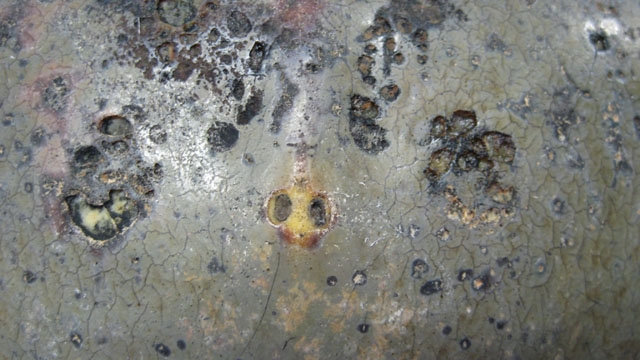
Gross Description:
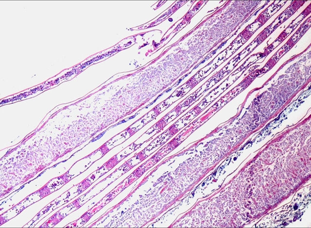
Histopathologic Description:
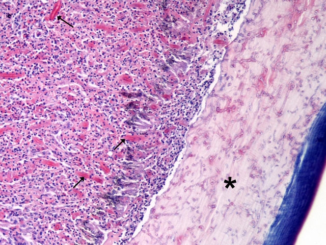
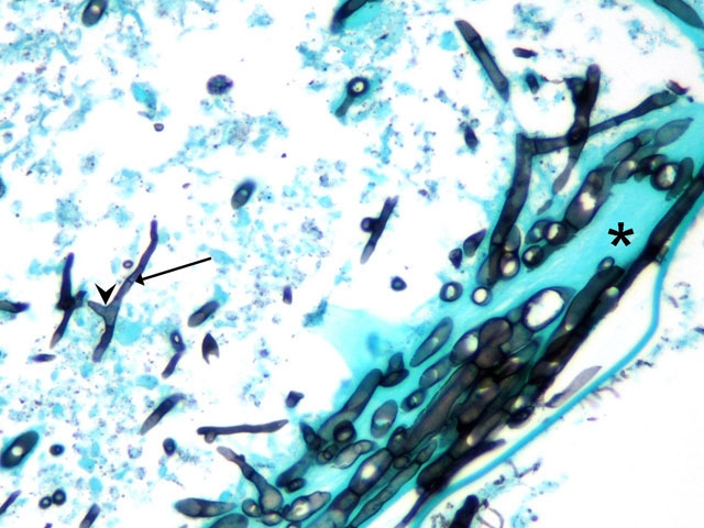
The cuticle of individual gill leaflets is multifocally thickened and penetrated by fungal hyphae. The central vascular channel of individual leaflets is expanded with hypereosinophilic and necrotic material, viable and degenerate amebocytes and bacteria (Fig. 3-3). There is necrotic debris mixed with bacteria between leaflets and sometimes pigmented fungal hyphae. Occasionally, there are some fungal hyphae on the surface of affected gill leaflets that are smaller (2-3 um diameter) than the invasive fungi.
Slides occasionally have metazoan parasites within the parenchyma and degenerate parasites associated with the surface of the carapace and/or gill leaflets.
Morphologic Diagnosis:
Gills: Branchitis, acute and chronic, necrotizing, multifocal, severe, with intralesional fungal hyphae and bacteria
Lab Results:
Condition:
Contributor Comment:
Mycotic shell disease has been reported only in captive horseshoe crabs. Specifically, mycotic infections of the carapace are reported in juvenile horseshoe crabs and most often in those housed without a sand substrate.(8,9) Gill and shell lesions similar to the ones seen in this crab are reported in a group of horseshoe crabs in a touch-tank at Ripley's Aquarium of the Smokies.(1) Single dose itraconazole therapy is well tolerated by horseshoe crabs, (1) but the efficacy of this treatment for the fungal lesions has not been investigated.
Fusarium species are saprophytes found ubiquitously within the environment and are the cause of diseases of both plants and animals including humans. Fusarium can cause disease in animals both by ingestion of mycotoxins and by fungal invasion of body tissues. Several examples of diseases caused by Fusarium infection in humans include keratitis, onychomycosis, dermatitis and disseminated disease.(3) Two recent reports of disease caused by Fusarium species reported in veterinary species include keratitis in a Holstein cow and intracranial fusariosis in a German Shepherd Dog.(4,5)
Invertebrate animals lack an adaptive immune system and respond to microbial antigens with a variety of innate immune responses including hemolymph coagulation, toll-like receptor mediated antimicrobial peptide production, melanin formation and lectin-mediated complement fixation. In horseshoe crabs, the hemolymph contains soluble antimicrobial proteins including C-reactive protein, alpha-2 microglobulins, lectins and hemocyanins. The granular amebocytes (also called hemocytes), which make up more than 99% of the circulating cells in the hemolymph, also contain antimicrobial proteins and coagulation proteins. Exposure to microbes causes degranulation of amebocytes and formation of a hemolymph clot. (6)
JPC Diagnosis:
2. Gills: Branchitis, necrotizing, acute and chronic, multifocal, severe, with fungal hyphae and bacteria
Conference Comment:
Morphologically, the Fusarium sp. are identified by hyaline, septate hyphae measuring 4 to 7um in width with frequent, usually right angle branching. This is important in helping to distinguish these from Aspergillus sp, which tend to have dichotomous branching and are 3-5um in width.(2)
References:
2. Chandler FW, Kaplan W, Ajello L: Color Atlas and Text of the Histopathology of Mycotic Diseases, pp. 76,79,101-102. Year Book Medical Publishers, Chicago, IL, 1980
3. Dignani MC, Anaissie E: Human fusariosis. Clin Microbiol Infect 10 (Suppl 1):67-75, 2004
4. Elligott CR, Wilkie DA, Kuonen VJ, Bras ID, Neihaus A: Primary Aspergillus and Fusarium keratitis in a Holstein cow. Vet Ophthalmol 9:175-178, 2006
5. Evans J, Levesque D, de Lahunta A, Jensen HE: Intracranial fusariosis: a novel cause of fungal meningoencephalitis in a dog. Vet Pathol 41:510-514, 2004
6. Iwanaga S, Lee BL: Recent Advances in the Innate Immunity of Invertebrate Animals. J Biochem Mol Biol 38:128-150, 2005
7. Maxie MG, Youssef S: In: Jubb, Kennedy and Palmers Pathology of Domestic Animals, ed. Maxie MG, 5th ed., pp. 358-359. Elsevier Limited, Philadelphia, PA, 2007
8. Smith SA: Horseshoe Crabs. In: Invertebrate Medicine, ed. Lewbart GA, 1st ed., pp. 133-142. Blackwell Publishing, Ames, IA, 2006
9. Smith SA, Berkson J: Laboratory culture and maintenance of the horseshoe crab (Limulus polyphemus). Lab Animal 34:27-34, 2005
10. Stalker MJ, Hayes MA: Liver and biliary system. In: Jubb, Kennedy and Palmers Pathology of Domestic Animals, ed. Maxie MG, 5th ed., pp. 371-372. Elsevier Limited, Philadelphia, PA, 2007