Wednesday Slide Conference, 2025-2026, Conference 4, Case 2
Signalment:
12-year-old, female, mixed breed, domestic goat, Capra aegagrus hircusHistory:
The owners report weight loss and cough for a few weeks. When auscultated, muffled heart sounds were heard on the right side, but no heart sounds were heard on the left side. Crackling sounds were heard in the dorsal lung fields but lungs sounds were absent ventrally. The owners elected for euthanasia and the goat was sent for a postmortem.Gross Pathology:
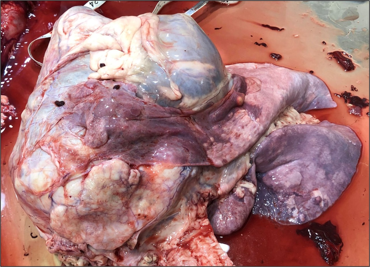
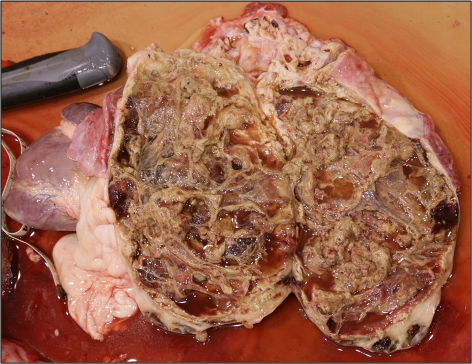
The goat was thin with small visceral and subcutaneous body fat stores. Moderate clear edema expanded the subcutis and deeper soft tissues along the ventral aspect of the mandibles, the ventral neck and the ventral trunk (dependent edema). Approximately 500 mls of orange-tinged fluid containing a few strands of fibrin was present in both the thoracic and abdominal cavities. Approximately 500 mls of similar fluid also distended the pericardial sac. An approximately 24 cm in greatest diameter, mass expanded the cranial mediastinum and filled the cranioventral thorax, displacing and compressing the lungs caudodorsally. When sectioned, much of the center of this mass was brown, necrotic and composed of many cystic pockets filled with yellow-brown clear fluid intermingled with pale yellow-tan, friable, debris. A thin peripheral layer of relatively intact tissue was generally present just beneath the variably thick layer of dense fibrous tissue which surrounded the mass. Portions of the cranioventral lung lobes were attached by extensive fibrous pleural adhesions to the outer surface of the mass. The pulmonary parenchyma was pale pink, partially collapsed and contained numerous, firm, randomly scattered, nodules ranging in size from 0.7-2.5 cm in greatest diameter. The base of the pulmonary trunk was markedly dilated just proximal to where it entered and passed through the large mediastinal mass. The right ventricular lumen and the right atrium were also markedly dilated. Mediastinal lymph nodes were unremarkable. The liver was diffusely dark red, moderately enlarged and when sectioned had a prominent red brown to tan, reticular (or nutmeg) pattern.Laboratory Results:
Immunohistochemistry on sections of the mediastinal mass was performed; see microscopic description.Microscopic Description:
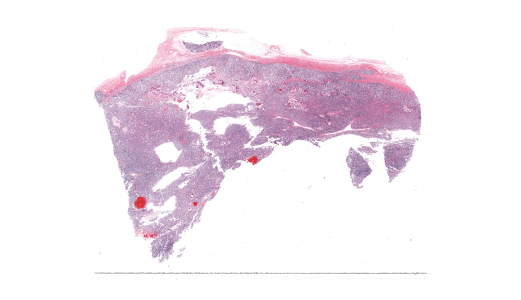
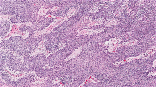
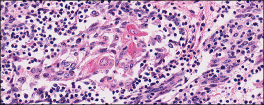
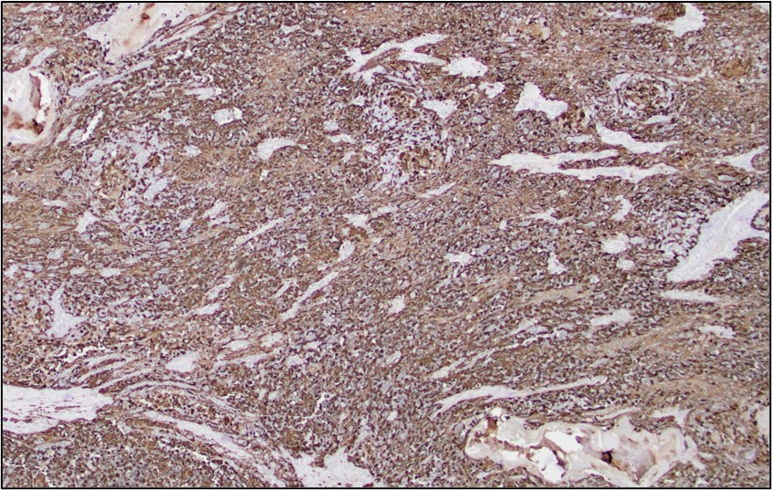
Much of the center of this mediastinal mass is effaced by extensive areas of necrosis and cystic degeneration. Sections were taken from the remaining thin peripheral rim of intact tissue that is surrounded by a variably thick, dense fibrous capsule. Neoplastic tissue is composed of highly cellular, infiltrates of plump spindloid to polygonal cells which are arranged in coalescing trabeculae and sheets and are intermingled with moderate to large numbers of small lymphocytes. These infiltrates multifocally infiltrate the surrounding capsule and are supported by small amounts of fine fibrovascular stroma which often forms dissecting thick bands and thinner septa. Frequently scattered within these infiltrates are small clusters of pale, polygonal, epithelial cells with glassy, keratinized, cytoplasm that are sometimes partially mineralized (Hassall’s corpuscles). The interstitium within the mass is also multifocally, mildly to moderately, expanded by pale poorly cellular fine fibrous stroma which often contains variably sized, deposits of basophilic, granular mineral. The majority of neoplastic polygonal to spindloid cells exhibit strong cytoplasmic staining with cytokeratin. Most of the small lymphocytes scattered within the mass were CD3 positive.The masses noted in the lung consist of similar, nodular mixed infiltrates of pale polygonal to spindloid cells intermingled with numerous small lymphocytes. These mixed infiltrates often dilated and fill pulmonary arteries and veins. Frequent scattered small deposits of mineral, foci of lytic and coagulative necrosis are also often scattered within these metastatic nodules.
Contributor's Morphologic Diagnoses:
Malignant thymoma/thymic carcinoma with pulmonary metastasisContributor's Comment:
In general, thymic epithelial tumors are considered uncommon tumors in veterinary medicine and have been reported humans and in a wide range of animals including, but not limited to, dogs, cats, rabbits, cattle, horses and sheep. The exception appears to be in older goats where, although neoplasia is generally uncommon, thymoma was reported as the third most common tumor in a retrospective study involving 100 goats5. One report involving a herd of dairy goats (mainly of Saanens or Saanen mixed breeds) found thymomas in 17 of 67 (25.3%) necropsied goats over 2 years of age2. Other reports have suggested that pygmy goats may also have a high incidence of thymomas3.In goats, thymomas are often considered benign, slow growing, tumors and many are incidental findings at necropsy or slaughter of mature animals. In most species, including goats, thymomas largely reside in the cranial mediastinum. However, due to the anatomy and development of the thymus in ruminants, whereby portions of this large lobulated organ in young animals may extend from the larynx to the cranial aspect of the pericardium, thymomas may form in ventral cervical region, the mediastinum, or involve both locations4. When large, as in this case, mediastinal thymomas may result in clinical signs such as respiratory distress and coughing due to compression and displacement of the lung, and trachea. Dependent edema in this goat was attributed to right heart failure caused by compression of the pulmonary trunk as it passed through this large mediastinal mass.
Histologically, thymomas can vary greatly in appearance and in composition. Neoplastic cells are epithelial and originated from endoderm of the third pharyngeal pouch in the fetus2. Tumor cells may appear polygonal and/or spindloid, and are accompanied by variable, sometimes large, numbers of mature, non-neoplastic lymphocytes. As in this case, neoplastic cells exhibit cytoplasmic staining with cytokeratins and most of the small lymphocytes present within the tumor are small CD3+ T cells. Thymomas have been sub-categorized, based on cellular composition as lymphocyte predominant, epithelial predominate, or mixed. A variety of sub-types have also been identified (clear cell, spindle cell, pigmented)8. A clear association with prognosis and these sub-types has not been identified in veterinary medicine. In human medicine, the 2004 World Health Organization (WHO) classification scheme of thymic epithelial tumors was created to better correlate histologic findings with the clinical behavior of these tumors. This system is also based on the cell morphology of neoplastic epithelial cells and the relative proportion of non-neoplastic lymphocytes present and appears to be applicable to canine thymomas9.
Malignant thymomas, or thymic carcinomas, are very rare but have been reported in a variety of species, including goats6. As in this case, the previously reported malignant thymoma involved a very large thymoma that had likely been present for a prolonged period and had many small pulmonary metastases. Thoracic lymph nodes were unaffected in the reported case, and in our goat.
A high incidence of paraneoplastic conditions is reported in association with thymomas in both human and veterinary medicine. In dogs, hypercalcemia due to production of PTH-rP is commonly associated with thymomas8. Autoimmune paraneoplastic conditions are also commonly associated with thymomas. Most thymomas exhibit varying, and sometimes marked, degrees of lymphopoiesis resulting in the proliferation of polyclonal CD4+ and CD8+ T cells. Normally in humans and animals, naïve T lymphocytes in the thymus interact with thymic epithelial cells which express high levels of MHC I and II molecules. This interaction results in the selection and weeding out of auto-reactive T cells that recognize both MHC molecules and self-antigens2. In animals with thymomas, aberrant expression by neoplastic epithelial cells of MHC molecules, autoantigens, and other critical receptors in this selection process, is thought to allow release of CD4+ and CD8+ T cells into systemic circulation which may then react with a wide variety of self-antigens. CD4+ T lymphocytes that recognize self-antigens (such as acetylcholine receptors) stimulate proliferation and differentiation of B lymphocytes with subsequent autoantibody production (resulting in myasthenia gravis and megaesophagus). In some conditions, such as thymoma-associated exfoliative dermatitis (which shares many clinical and histologic features with erythema multiforme), autoreactive lymphocytes bind directly to keratinocytes and induce apoptosis1. A wide variety of additional autoimmune diseases have been reported in animals in association with thymomas including polymyositis, thrombocytopenia, anemia, and granulocytopenia8. Paraneoplastic disease in goats with thymomas is extremely rare but megaesophagus and ruminal tympany attributed to esophageal compression by a large mediastinal mass, and thymoma-associated exfoliative dermatitis have both been described in domestic goats 1,7.
Contributing Institution:
Atlantic Veterinary College, University of Prince Edward Island www.upei.ca/avcJPC Diagnoses:
Thymus: Thymoma.JPC Comment:
The contributor provided an excellent, thorough explanation of thymomas, the paraneoplastic syndromes seen with this neoplasm, and the pathogeneses of how thymomas cause these syndromes. These topics were central to the conference discussion of this case, as well as to the types of questions that were asked of participants about thymomas. As such, the contributor’s comment is well-worth a complete read.Another topic expounded upon was of the differing types of thymomas classified in human patients and how applicable these are to veterinary medicine. The major thymoma classifications according to the World Health Organization are Types A, A/B, B1, B2, and B3.6 Type A thymomas are defined by their predominance of spindle-shaped epithelial cells and lack of T-cells.6 Type A/Bs, also known as “mixed” thymomas, have similar epithelial cell morphology but have an abundance of T-cells throughout.6 Type B1 has abundant immature T-cells, areas of medullary differentiation, and few epithelial cells.6 Type B2 is described as having abundant immature T-cells with greater numbers of singular or clustered epithelial cells dispersed throughout the lymphocytes.6 Lastly, Type B3 has sheets of epithelial cells with rare intercellular bridging and a paucity of T-cells.6
Thymoma classification has been discussed in dogs, goats, and sheep predominantly, and there seems to be a shift towards trying to align thymoma diagnoses in veterinary species with the classification system that the WHO utilizes for humans. Although it has yet to be fully elucidated whether these classifications are truly comparable in veterinary medicine, current literature states that approximately 15% of canine thymomas, as well as the overwhelming majority of thymomas in goats and sheep, are most consistent with Type A/B, although other types have been reported.10
References:
- Byas AD, Applegate TJ, Stuart A, Byers S, Frank CB. Thymoma-associated exfoliative dermatitis in a goat: case report and brief literature review. J Vet Diagn Invest, 2019;31(6):905-908.
- Boes KM, Durham AC. Bone marrow, blood cells and the lymphoid/lymphatic system. In: Zachery JF, ed. Pathologic Basis of Veterinary Disease, 6th St’ Louis, Missouri: Elsevier; 2017: 761-764.
- Hadlow WJ. High Prevalence of thymoma in the dairy goat: report of 17 cases. Vet Pathol, 1978;15:153-169.
- Hill JA, Fubini SL, Hackett RP. Clinical features, treatment, and outcome in goats with thymomas: 13 cases (1990-2014). JAVMA; 2017;251(7):829-834.
- Lohr CV. One hundred two tumors in 100 goats. Vet Pathol, 2012; 50(4):668-675.
- Marx A, Chan JKC, Chalabreysse L, Dacic S, Detterbeck F, French CA, Hornick JL, Inagaki H, Jain D, Lazar AJ, Marino M, Marom EM, Moreira AL, Nicholson AG, Noguchi M, Nonaka D, Papotti MG, Porubsky S, Sholl LM, Tateyama H, Thomas de Montpréville V, Travis WD, Rajan A, Roden AC, Ströbel P. The 2021 WHO Classification of Tumors of the Thymus and Mediastinum: What Is New in Thymic Epithelial, Germ Cell, and Mesenchymal Tumors? J Thorac Oncol. 2022;17(2):200-213.
- Olchowy TWJ, Toal RL, Brenneman KA, Slauson DO, McEntee MF. Metastatic thymoma in a goat. Can Vet J, 1996; 37:165-167.
- Parish SM, Middleton JR, Baldwin TJ. Clinical megaesophagus in a goat with thymoma. Vet Rec, 1996; 139:94.
- Valli VE, Bienzle D, Meuton DJ. Tumors of the Hemolymphatic System. In: Meuton DJ, ed. Tumors in Domestic Animals, 5th Ames, Iowa; Wiley Blackwell; 2017: 305-307.
- Valli VEO, Kiupel M, Bienzle, D, Wood RD. Hematopoietic System. In: Maxie MG, ed. Jubb, Kennedy, and Palmer’s Pathology of Domestic Animals, 6th St’ Louis, Missouri: Elsevier; 2016: Vol 3; 150-158.