Signalment:
Gross Description:
Histopathologic Description:
Small intestine (not submitted): In the villus epithelium, meronts containing merozoites identical to those observed in the liver are abundant. Release of the merozoites into the lumen is also observed.
Morphologic Diagnosis:
Condition:
Contributor Comment:
JPC Diagnosis:
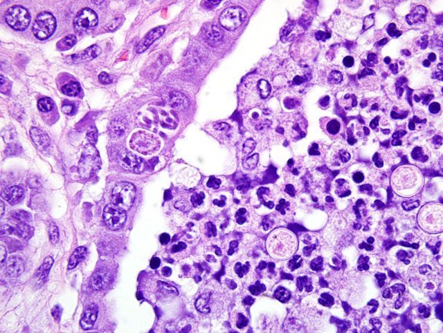
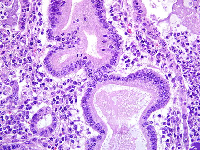
1. Liver, biliary tract: Cholangitis and pericholangitis, chronic, neutrophilic and lymphoplasmacytic, diffuse, moderate to marked, with biliary hyperplasia, and numerous intraepithelial apicomplexan schizonts and gametes, and intraluminal apicomplexan oocysts.
2. Liver: Extramedullary hematopoiesis, multifocal, marked.
Conference Comment:
| Eimeria and Isospora of Animals | ||
|---|---|---|
| Host | Coccidian | Organ(s) Affected |
| Cattle | E. zuernii E. bovis | Distal small intestine Distal small intestine |
| Sheep | E. ovinoidalis E. ashata E. bakuensis E. crandallis | Terminal ileum/ cecum and colon Distal small intestine Distal small intestine Distal small intestine |
| Goats | E. ninakohlyakimovae E. caprina E. christenseni | Cecum and colon Cecum and colon Distal small intestine |
| Pigs | Isospora suis (neonatal pigs) E. scabra (weaners, growers) E. debliecki (weaners, growers) E. spinosa (weaners, growers) | Distal small intestine Distal small intestine Distal small intestine Distal small intestine |
| Horses | E. leuckarti | Small intestine |
| Dogs | C. canis C. ohioensis complex = (C. burrowsi, C. ohioensis, C. neorivolta) | Distal small intestine, large intestine Distal small intestine, large intestine |
| Cats | C. felis C. rivolta | Small intestine, large intestine Small intestine, large intestine |
| Mice | E. falciformis | Colon |
| Rabbits | E. stiedae E. intestinalis E. flavescens | Bile ducts Ileum, cecum Ileum, cecum |
| Guinea Pig | E. caviae | Large intestine |
| Ferret | E. furonis1,4 E. ictidea1,4 | Small intestine, Bile ducts Small intestine |
| Chickens | E. acervulina E. necatrix E. tenella | Small intestine Small intestine Cecum |
Morphologic features used to distinguish the various coccidia, such as Cryptosporidium, Besnoitia, Sarcocystis, Toxoplasma, Eimeria, and Isospora were also discussed. The primary characteristic used to differentiate the various genera of coccidia is the structure of the sporulated oocyst, particularly the number of sporocysts and sporozoites present, as summarized below.(2)
| Apicomplexa Sporocyst and Sporozoite Numbers | ||
|---|---|---|
| Coccidian | Sporocysts | Sporozoites |
| Cryptosporidium | 0 | 4 |
| Besnoitia Isospora Sarcocystis Toxplasma | 2 | 8 |
| Eimeria | 4 | 8 |
Additional differentiating characteristics, such as the host affected, location of the parasite within the host, and organism size and shape, were also mentioned as criteria to be considered when speciating coccidia. The moderator noted that Eimeria and Isospora may be also differentiated by their location of replication; Eimeria spp. replicate within the epithelium while Isospora spp. replicate within the lamina propria.
Attendees briefly reviewed the coccidian life cycle using E. stiedae as an example. In short, oocysts are shed in feces and sporulate. Sporulated oocysts contain four sporozoites that hatch within the intestine and then enter the liver via the portal vein. Sporozoites then penetrate into the bile epithelium and form trophozoites that undergo asexual nuclear division (schizogony). Schizogony results in the formation of schizonts that contain merozoites. The schizonts rupture, damaging the cell, and release merozoites which infect additional cells. Eventually, merozoites form sexual stages (i.e. male microgametes and female macrogametes) which unite to form oocysts. Oocysts are released into the bile and shed into the feces and the cycle then repeats itself.
Participants discussed the similarities and dissimilarities between the coccidian in this case and Eimeria stiedae. Both replicate within the hepatobiliary epithelium and cause chronic cholangiohepatitis, with marked epithelial hyperplasia, bile duct reduplication, and portal fibrosis.(3) Additionally, E. stiedae produces long papillary fronds within the bile duct that are not present in the case of this ferret. The moderator noted that hepatobiliary coccidiosis is rare in ferrets and has only been reported in very young animals that are generally less than four months of age. The vacuolation of hepatocytes noted in this case by attendees is a very common finding in ferrets due to inanition and fatty mobilization.
References:
2. Gardiner CH, Fayer R, Dubey JP: An Atlas of Protozoan Parasites in Animal Tissues, 2ne ed., p. 20, Armed Forces Institute of Pathology, Washington, D.C., 1998
3. Thompson, ME: Proceedings: Department of Veterinary Pathology Wednesday Slide Conference 2006-2007. Armed Forces Institute of Pathology, Washington, D.C., 2007
4. Williams BH, Chimes MJ, Gardiner CH: Biliary coccidiosis in a ferret (Mustela putorius furo). Vet Pathol 33:437-439, 1996