Signalment:
15-year-old, female, Manx cat,
Felis domesticusTwo liver masses (4-6 cm in diameter) were discovered during a regular visit to the local vet. The cat was clinically healthy.
Histopathologic Description:
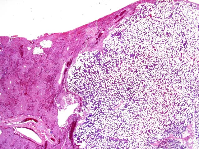
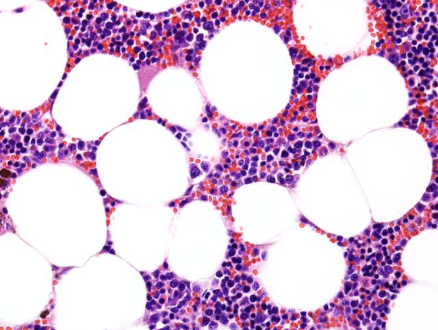
Sections of liver were partially effaced by a well-demarcated and unencapsulated mass composed of histologically normal adipose tissue and different cells of hematopoietic series. Cellular atypia and mitoses were not significant.
Morphologic Diagnosis:
Myelolipoma, liver
Condition:
Myelolipoma, multiple
Contributor Comment:
Myelolipomas are rare, benign, and endocrinologically inactive tumors composed histologically of adipocytes with variable hematopoietic cells, including both mature and immature cells of the granulocytic, erythrocytic, and megakaryocytic series. They have been reported rarely in veterinary literature, particularly in the liver of cats and wild felidae; and in the spleen, adrenal gland, and spinal cord of dogs.(1,2) The vast majority of myelolipomas in humans occur within the adrenal glands, but extra-adrenal myelolipomas have been reported.(1) Feline hepatic myelolipomas are usually of no clinical significance, and they are usually diagnosed incidentally at necropsy during exploratory laparotomy or during abdominal ultrasonography. Metastasis has not been reported.(2) The pathogenesis of myelolipoma remains speculative. Theories concerning the pathogenesis include autonomous proliferation of bone marrow cells transferred during embryogenesis or metaplasia of certain mesenchymal cells triggered by various stimuli, including necrosis, infection, or stress.
When considering the differential diagnosis of hepatic myelolipomas, extramedullary proliferations of hematopoietic elements (EMH) is the primary differential. Myelolipomas are welldemarcated, discrete, single or multiple masses in any part of the liver; whereas, EMH appears as multifocal microscopic aggregates of hematopoietic cells in the perisinusoidal compartment and in portal areas. Also, hepatic EMH is usually not associated with forming discrete masses; however, severe EMH can lead to diffuse organ enlargement (hepatomegaly).
JPC Diagnosis:
Liver: Myelolipoma, multiple
Conference Comment:
Myelolipomas have also been found in the adrenal glands of cattle, non-human primates, and sporadically in other various species. Histologically, these benign lesions are similar to those in the liver and are composed of both myeloid and erythroid hematopoietic tissue admixed with well-differentiated adipocytes. Bone formation and areas of mineralization have also been observed in these tumors. Rare cases of splenic myelolipomas have also been reported. Grossly, these lesions stand out from the splenic parenchyma as a focal area of pallor. The speculative pathogenesis of this lesion is metaplastic transformation of a resident cell population regardless of location (adrenal gland, liver, and spleen).(3)
References:
1. Al-Rukibat RK, Bani Ismail ZA: Unusual presentation of splenic myelolipoma in a dog. Can Vet J. 47(11):1112-4. 2006
2. Capen CC: Endocrine glands.
In: Jubb, Kennedy, and Palmers Pathology of Domestic Animals, ed. Maxie MG, 5th ed., vol. 3, pp.415. Elsevier Limited, St. Louis, MO, 2007
3. Stalker MJ, Hayes MA: Liver and biliary system.
In: Jubb, Kennedy, and Palmers Pathology of Domestic Animals, ed. Maxie MG, 5th ed., vol. 2, pp. 386. Elsevier Limited, St. Louis, MO, 2007
4. Valli VEO: Hematopoietic system.
In: Jubb, Kennedy, and Palmers Pathology of Domestic Animals, ed. Maxie MG, 5th ed., vol. 3, pp. 289. Elsevier Limited, St. Louis, MO, 2007