Signalment:
28-year-old, male, American alligator, (
Alligator mississippiensis).This
alligator was housed with 10 others at a rescue facility in the northeastern
United States. During the winter months, the animals were kept indoors in a
house, living on wood floors, with access to an unspecified water source; there
was reportedly visible mold in this environment. The alligators were not
induced to hibernate during this time, and were housed at an ambient
temperature of 74
oF (23
oC). This alligator was found dead
with no premonitory signs.
Gross Description:
An
adult, 70.5 kg male American alligator was presented in good nutritional
condition with abundant fat stores.
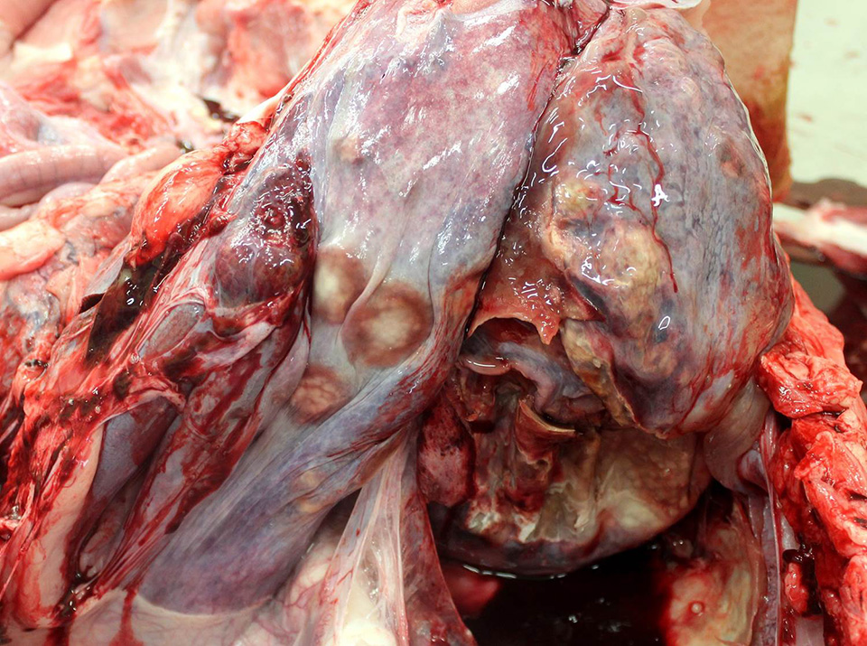
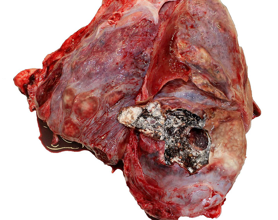
There was an extensive adhesion of
the right lung to the right dorsal body wall which enclosed approximately 75 mL
of pale tan, thin liquid with grey to pale tan particulates. The pleural
surfaces of both lungs had discrete to coalescing areas of firm, brown to pale
tan discoloration which were more extensive in the right lung and extended into
the parenchyma. On cut section there were extensive regions of parenchyma
replaced by firm, white, crumbly material (caseous necrosis). Additionally,
multiple subpleural cavitations and air spaces in the right lung were lined by
white, slightly fuzzy, fungal mats. Numerous small, whitish foci up to 0.3 cm
diameter were present in the liver, and similar individual foci were present in
the heart, spleen, and kidney.
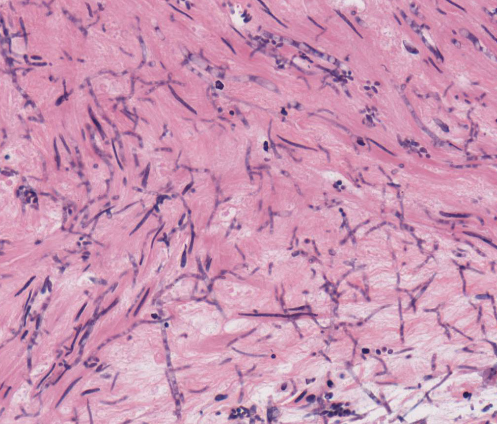
Histopathologic Description:
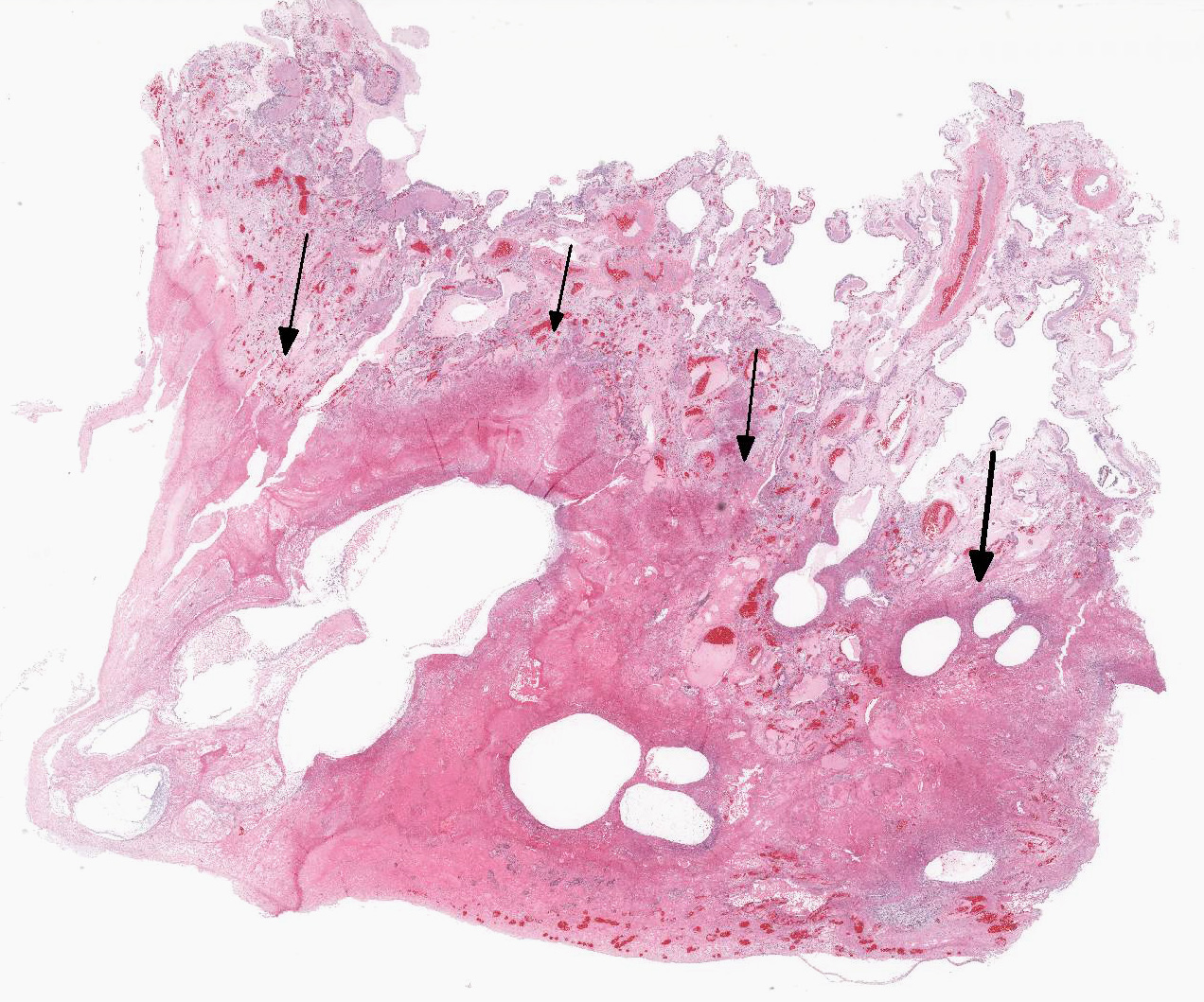
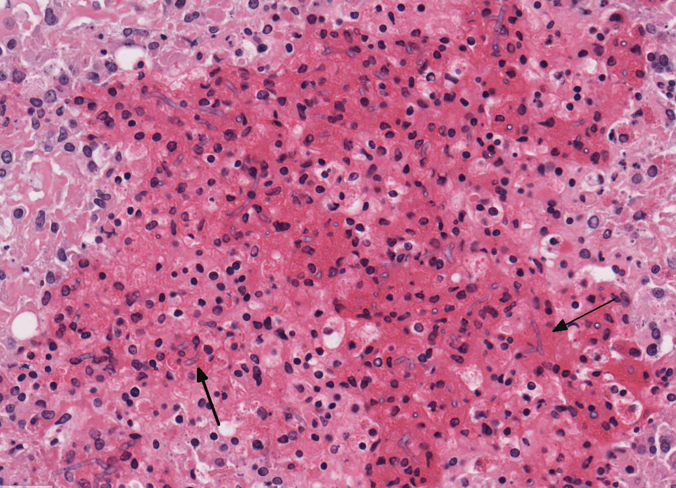
Up
to 80% of the parenchyma is effaced by necrosis that extends to the pleural
surface. Necrotic areas are characterized by variable loss of architectural detail
with accumulation of fibrin, hypereosinophilic cellular debris, edema,
hemorrhage, and inflammatory cells composed predominantly of heterophils and
macrophages. Within the necrotic areas, there are numerous, lightly basophilic
to transparent fungal hyphae that are 2-6 microns in diameter, septate, and
parallel-walled with occasional right angle branching. At the air-tissue
interface in a couple of large airways there are scattered hyphae that give
rise to dense clusters of ampulliform conidiogenous cells measuring up to 3
microns in diameter, each with a single to several terminal round conidia
measuring 1-3 microns in diameter. Large numbers of bacteria are also
frequently admixed. Rarely, in areas of dense fungal growth, there are a few
translucent, variably shaped, anisotropic crystals (oxalate crystals).
Frequently, vessels are occluded by fibrin thrombi, and their walls contain
necrotic debris, fibrin, moderate numbers of degenerate heterophils and
occasional fungal hyphae (vasculitis). The parenchyma adjacent to necrotic
areas is variably expanded by edema, mixed inflammatory cells, and reactive
fibroblasts.
Morphologic Diagnosis:
Lung: severe,
subacute, multifocal to coalescing, fibrinonecrotizing and hetero-philic
pneumonia and pleuritis with vasculitis, fibrin thrombi and intralesional
bacteria and fungal hyphae and conidia, consistent with
Beauveria bassiana.
Lab Results:
Fungal
culture and identification: A swab from the lung was initially plated on
potato dextrose agar (PDA) and inhibitory mold agar (IMA) with and without
antibiotics and then incubated at 30
oC for 14 days; this yielded
heavy growth of white fungal colonies on both plate types. On PDA, the bottom
of the colony had an orange to pink tinge, whereas on IMA the bottom of the
colony was red. Cultures were submitted to the Fungus Testing Laboratory,
University of Texas Health Science Center in San Antonio, Texas for
identification. Combined phenotypic characterization and DNA sequencing of ITS
and TEF targets identified the fungus as
Beauveria bassiana.
Condition:
Beauveria bassiana, alligator
Contributor Comment:
This
is a case of mycotic pneumonia in an American alligator caused by
Beauveria
bassiana.
Identification of this organism was based on the morphology
of the fruiting bodies (conidiogenous cells and conidia) on H&E; its
phenotypic characteristics in culture; and DNA sequence analysis, all of which
differentiated it from other common agents of fungal pneumonia, particularly
Asper-gillus
species.
Beauveria bassiana is a ubiquitous soil saprophyte that is
entomopathogenic, i.e. pathogenic to insects due to an affinity for chitinous
exoskeletons. As such, it has been widely used for more than 100 years as
biocontrol of pest insects.
5 Though widespread in the environment,
its upper temperature limit is around 30
oC, and it is inactivated
within hours or days when exposed to sunlight.
9 Due to the
temperature limitations,
B. bassiana rarely causes infections in mammals
but is an opportunistic pathogen of reptiles, with previous reports in captive
American alligators,
2 chelonians,
3,6 and in cold-stunned
Kemps Ridley sea turtles.
62 In the current case, the temperature
of the indoor enclosure was reportedly kept at 23
oC, well within the
temperature range of
B. bassiana. High levels of fungus in the
environment and poor ventilation were also probably involved in this case, as
mold was reportedly visible in the enclosure where this group of alligators was
housed. Other predisposing factors for fungal pneumonia in captive reptiles
include additional husbandry-related issues, such as humidity, hygiene, and
nutrition, immunosuppression, overuse of antibiotics, and concurrent disease.
5
Shortly after diagnosis of this case, a second alligator from the same group
died naturally, but a necropsy was not performed. Transmission is
thought to occur from inhalation or ingestion of fungal spores from the
environment, and the lung appears to be the primary site of infection.
Hematogenous dissemination of the infection from the lung to other tissues,
such as liver and spleen, occurred in this case as in previous cases.
2,6
Beauveria bassiana produces several toxic compounds including oxalic
acid, which promotes the formation of oxalate crystals within affected tissues;
9
only a few crystals were seen in this case.
JPC Diagnosis:
Lung: Pneumonia, necrotizing, multifocal to coalescing, severe,
with innumerable fungal hyphae and large colonies of mixed bacilli, American
alligator,
Alligator mississippiensis.
Conference Comment:
This
case provided conference participants the unique opportunity to describe lung
pathology in an American alligator, an uncommonly seen species at the Joint
Pathology Center. Prior to the discussion of this case, the conference
moderator led a review of the normal functional anatomy and physiology of
alligator lungs, which was poorly understood until relatively recently.
1,7
Research performed at the University of Utah indicates the external and
internal morphology of alligator lungs is strikingly similar to the avian
respiratory system, although in contrast to birds, alligators lack
intra-abdominal air sacss.
1,7 Alligators have a highly efficient
unidirectional style of breathing, originally thought to be unique to avian
species as a consequence of the high oxygen demands of flight.
7
However, unlike birds, alligators use a diaphragm to pull air into the lungs.
The air then travels one direction through bronchi which branch into numerous
smaller parabronchi and continues further into alveolar-like spaces, called
faveoli. Gas exchange then takes place within these faveoli, and the air then
flows out of the lung via in a one-way loop and valve system.
7
Unidirectional breathing is much more efficient than the mammalian
bellows-style breathing because there is no alveolar mixing of inspired and
expired air. Research is ongoing to elucidate the exact mechanism of
unidirectional air flow in alligators and other reptiles, as it was thought
that air sacs were necessary for unidirectional air flow breathing.
1,7
Reported cases of fungal
pneumonia in reptiles caused by
Beauveria bassiana are rare and
typically involve extensive multifocal necrosis or granulomatous nodules with
high numbers of fungal hyphae in the lungs with dissemination to the multiple
abdominal organs, as present in this case.
2,3,6 Infection occurs
after inhaling or ingesting fungal spores from the environment and development
of disease in reptiles has been associated with low environmental temperatures
and poor husbandry of captive reptiles. As mentioned by the contributor, the fungus will not grow at mammalian
physiologic temperatures (37)
2,3,6,
although it has been very rarely reported to cause fungal keratitis in people
associated with contact lens wear and prior treatment with corticosteroid eye
drops.
4 The association of this fungus with low environmental
temperatures and cold-shocked reptiles in previously reported cases prompted
the conference moderator to discuss brumation in ectothermic animals. Brumation is a time of
dormancy in reptiles in response to colder winter weather (~21
oC),
and is similar, but not identical, to hibernation in mammals. During periods of
brumation, reptiles have a markedly decreased metabolic rate, but do not fall
into a deep sleep, and can regularly emerge to drink and bask during warm days.
Additionally, reptiles typically do not eat during periods of brumation.
8
References:
1. Farmer
CG. Similarity of Crocodilian and avian lungs indicates unidirectional flow is
ancestral for Archosaurs.
Integr Comp Biol. 2015; 55(6):962-971.
2. Fromtling
RA, Jensen JM, Robinson BE, Bulmer GS. Fatal mycotic pulmonary disease of
captive American alligators.
Vet Pathol. 1979; 16:428-431.
3. Gonzalez
Cabo JF, Espejo Serrano J, Barcena Asensio MC. Mycotic pulmonary disease by
Beauveria
bassiana in a captive tortoise.
Mycoses. 1995; 38:167-169.
4. Lara
Oya A, Medialdea ME, et al. Fungal keratitis due to
Beauveria bassiana
in contact lenses wearer and review of published reports.
Myco-pathologia.
2016; 181:745-752.
5. Murray
MJ. Pneumonia and normal respiratory function. IN Mader, DR, ed.
Reptile Medicine
and Surgery, St. Louis: W.B. Saunders Company; 1996: 402-403.
6. Pare
JA, Jacobson ER. Mycotic diseases of reptiles. IN Jacobson ER, ed.
Infectious
Diseases and Pathology of Reptiles, Boca Raton: CRC Press; 2007: 538-539.
7. Sanders
RK, Farmer CG. The pulmonary anatomy of
Alligator mississippiensis and
its similarity to the avian respiratory system.
Anat Rec. 2013;
295(4):699-714.
8. >Wilkinson
A, Hloch A, et al. The effect of brumation on memory retention.
Sci Rep.
2017; 7:40079.
9. Zimmermann
G. Review on safety of the entomopathogenic fungi
Beauveria bassiana and
Beauveria brongniartii.
Biocontrol Sci Technol. 2007; 17(6):553-596.