CASE I:
Signalment:
15-month-old, female, French bulldog, Canislupus
History:
Fifteen month old French bulldog with chronic diarrhea for months that occasionally contained blood but not always. Some weight loss with a current BCS 4.5/10. Fecal smear was positive for giardia. Treated with fenbendazole / metronidazole. No more blood reported in stools but still chronic diarrhea. The dog was euthanized and the necropsy revealed very thickened large intestine and distal small intestine. Owner says they have had other dogs with similar symptoms without any successful treatment.
Laboratory Results:
No laboratory findings reported.
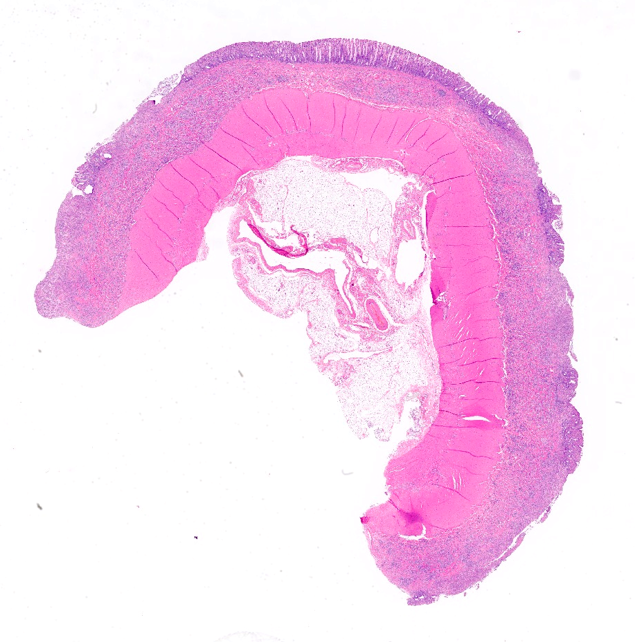
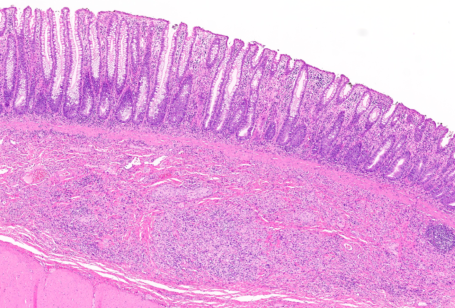
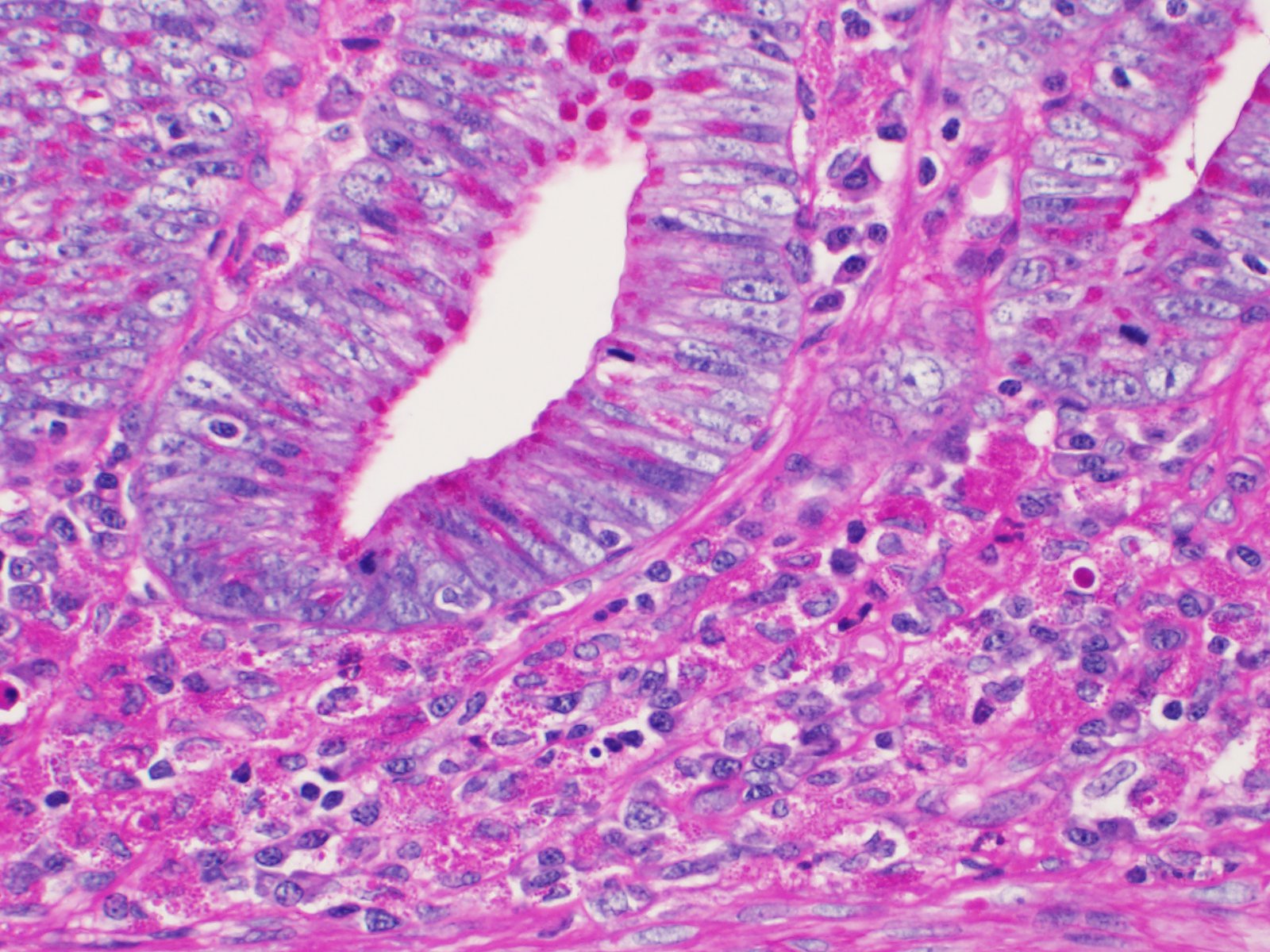
Microscopic Description:
Within the submitted and examined sections of colon the mucosa and underlying submucosa is markedly expanded by a dense infiltrate composed of large numbers of macrophages admixed with lesser numbers of neutrophils, lymphocytes, and plasma cells. These macrophages often contain abundant vacuolated cytoplasm with karyorrhectic debris and rare intracytoplasmic basophilic rod-shaped bacteria. There is also multifocal to coalescing ulceration of the mucosa that varies from superficial to nearly full thickness. The deep mucosa and submucosa underlying the most severely affected areas contains increased numbers of neutrophils. The colonic glands are mildly hyperplastic with evidence of regeneration including piling of glandular epithelium, karyomegaly, increased cytoplasmic basophilia, and frequent mitotic figures. Occasionally these glands contain small numbers of intraluminal spirochete-like organisms.
The macrophages present within the mucosa and submucosa contain frequent PAS positive material within their cytoplasm.
Contributor’s Morphologic Diagnoses:
Colon: Histiocytic and ulcerative colitis, multifocal to coalescing, severe
Contributor’s Comment:
Histiocytic ulcerative colitis (HUC), also known as granulomatous colitis (GC), is a chronic enteropathy of dogs that was first reported in 1965.15 It is histologically characterized by marked infiltration of the colonic mucosa and submucosa by large, periodic acid-Schiff (PAS)-positive macrophages; these are typically accompanied by mucosal ulceration, loss of goblet cells8, and infiltrates of lymphocytes, plasma cells, and neutrophils.1,13,15 Histopathological demonstration of macrophages with intracytoplasmic PAS positive material has been accepted as the pathognomonic feature of HUC4,10 and the PAS positive material is considered to be glycoprotein from bacterial cell walls.9
Although this condition has nonspecific gross lesions, findings in previously reported cases include reduction in the colonic length, eccentric thickening of the intestinal wall, and multifocal to coalescing areas of ulceration that can be present in the colon, cecum and rectum.2,4,14 Clinical features include chronic diarrhea, increased frequency of defecation, and feces that are soft, tan, granular, glistening, and sometimes or always contain blood. Affected dogs are afebrile and late in disease often develop dehydration with weight loss.16
HUC has been described most commonly in young Boxers and French Bulldogs, although it has been sporadically reported in a variety of other breeds; Mastiff, Alaskan Malamute, Doberman Pincher, American Staffordshire Terrier1, and Beagle.2,13,14 It has also rarely been reported in cats, including a Persian-crossbred and two domestic shorthaired cats.6,9,15
HUC was previously regarded as an idiopathic immune-mediated disease14, and although the exact cause and pathogenesis are still not well understood, recent reports have identified Escherichia coli in both canine1,2,5,7,8,11 and feline cases.6,9 In both species, clinical remission as well as histologic resolution has been described after eradication of the invasive E. coli by use of antibiotics including Enrofloxicin.8,9 The response to these treatments varies by case and antibacterial resistance, especially to enrofloxacin, can affect the outcome with nonresponsive or refractory cases.14 Thus, the regression of clinical and histologic lesions suggest that E. coli has a critical role in the development of HUC.9 The predisposition of Boxers to HUC is thought to be due to a heritable anomaly that confers susceptibility to the invasion and persistence of an adherent and invasive group of E. coli.1 The rarity of disease in cats may suggest variation in hereditary and host susceptibility to E coli-induced HUC between dogs and cats.9
Contributing Institution:
Kansas State Veterinary Diagnostic Laboratory (KSVDL)
http://ww.ksvdl.org/
JPCDiagnosis:
Colon: Colitis, histiocytic and ulcerative, diffuse, marked, with glandular hyperplasia.
JPC Comment:
While the presence of PAS-positive cytoplasmic material in macrophages on histopathology is virtually pathognomonic for histiocytic ulcerative colitis, fluorescent in-situ hybridization (FISH) is the definitive diagnostic test for identifying E. coli in this condition.3, 8, 12 This technique uses fluorescent probes to specifically target and hybridize with E. coli 16S ribosomal rDNA, thus enabling identification and localization of the bacteria within HUC lesions.8 In addition to allowing direct observation of E. coli within macrophages, FISH has several methodological advantages: it can be conducted on formalin-fixed paraffin-embedded sections, it is not confounded by normal flora present in the colon, and it can detect multiple bacterial species at once.12
Two recent reports have described the correlation of histologic and cytologic findings in two dogs with HUC.3, 12 Both dogs had a history of chronic diarrhea and hematochezia and were painful with thickened, irregular mucosa on rectal exam.3, 12 Rectal scrapings in both dogs revealed mixed inflammation and occasionally macrophages contained pink cytoplasmic granules or phagocytosed bacteria.3, 12 In one case, the pink cytoplasmic material was confirmed to be PAS positive.3 In both cases, HUC was confirmed with histopathology and FISH.3, 12 These are the first published reports of cytologic findings of HUC, so the sensitivity and specificity of rectal scrapings for this condition is unknown.12 The results suggest, however, that cytologic examination may be helpful as an initial test for guiding further diagnostics, particularly since it is non-invasive, economical for owners, and does not require a specialized laboratory for analysis.
References:
- Argenta FF, de Souza SO, Meirelles, LS, et al. Histiocytic ulcerative colitis in an American Staffordshire Terrier. J Comp Path. 2018;165:40-44.
- Carvallo FR, Kerlin R, Fredette C, et al. Histiocytic typhlocolitis in two colony Beagle dogs. Exp Toxicol Pathol 2015;67:219-221.
- Conrado FO, Jones EA, Graham EA, Simpson KW, Craft WF, Beatty SSK. Cytologic, histopathologic, and clinical features of granulomatous colitis in a French bulldog. Vet Clin Pathol. 2021;00:1-7.
- German AJ, Hall EJ, Kelly DF, Watson ADJ, Day MJ. An immunohistochemical study of histiocytic ulcerative colitis of Boxer dogs. J Comp Path. 2000;122:163-175.
- Hostutler RA, Luria BJ, Johnson SE, Weisbrode SE, Sherding RG, Jaeger JQ, et al. Antibiotic-responsive histiocytic ulcerative colitis in 9 dogs. J Vet Intern Med. 2004;18:499-504.
- Leal RO, Simpson K, Fine M, Husson J, Hernandez J. Granulomatous colitis: more than a canine disease? A case of Escherichia coli-associated granulomatous colitis in an adult cat. J Feline Med Surg. 2017;3:1-5.
- Manchester AC, Hill S, Sabatino B, et al. Association between granulomatous colitis in French Bulldogs and invasive Escherichia coli and response to fluoroquinolone antimicrobials. J Vet Intern Med 2013;27:56-61.
- Mansfield CS, James FE, Craven M, Davies DR, OHara AJ, Nicholls PK, et al. Remission of Histioytic ulcerative colitis in boxer dogs correlates with eradication of invasive intramucosal Escherichia coli. J Vet Intern Med.2009;23:964-969.
- Matsumoto I, Nakashima Ko, Morita H, Kasahara K, Kataoka O, Uchida K. Escherichia coli-induced granulomatous colitis in a cat. J Feline Med Surg. 2019;5:1-5.
- Nolte A, Junginger J, Baum B, et al. Heterogeneity of macrophages in canine histiocytic ulcerative colitis. Innate Immun 2017;23:228-239.
- Simpson KW, Dogan B, Rishniw M, et al. Adherent and invasive Escherichia coli is associated with granulomatous colitis in boxer dogs. Infect Immun 2006;74:4778-4792.
- Sims CS, Nagle J, Tolbert MK, Anderson K, Linder K, Neel J. Correlation of cytology to histology in a case of granulomatous colitis in a Boxer dog. Vet Clin Pathol. 2022:50(Suppl.1):83-87.
- Stokes JE, Kruger JM, Mullaney T, Holan K, Schall W. Histiocytic ulcerative colitis in three non-boxer dogs. J Am Anim Hosp Assoc.2001;37:461-465.
- Uzal FA, Plattner BL, Hostetter JM. Alimentary system. In: Maxie MG, ed. Jubb, Kennedy and Palmer’s Pathology of Domestic Animals. 2. 6th ed. Philadelphia, PA: Elsevier. 2016:97-98.
- Van Kruiningen HJ and Dobbins WO 3rd. Feline histiocytic colitis. A case report with electron microscopy. Vet Pathol. 1979;16:215-222.
- Van Kruiningen HJ, Montali RJ, Strandberg JD, Kirk RW. A granulomatous colitis of dogs with histologic resemblance to Whipple’s disease. Path Vet.1965;2:521-544.