WSC 2022-2023
Conference 19
Case III:
Signalment:
13 Years, male, indoor only cat, Felis catus
History:
The cat presented at the vet because the owner had realized that the right eye seemed “bigger”, accompanied by a widened pupil. The clinical examination revealed normal pulse, mucosal surfaces, abdomen and lymph nodes as well as nondescript auscultation of heart and lung.
The intraocular pressure in the right eye was 59 mm Hg initially, and the intraocular pressure was 25 mmHg in the left eye. After one week of treatment with Azopt three times per day, the intraocular pressure of the right eye was reduced to 21 mmHg at the second consultation.
Values of the clinical examination at the second consultation:
- Blood pressure: 160/120 (5 measurements)
- Glucose: 6.4 mmol/l
- T4: within the normal range
- Renal values: within the normal ranges
Clinical examination of the right eye: eye is open; reflexes are negative apart from a minimal pupil reflex; eye lids and their position are in within normal range; the conjunctiva is pink; the cornea is clear; the anterior eye chamber is clear; iris is in mydriasis; the lens demonstrates mild nucleosclerosis; the corpus vitreous with partly ablated retina in nasal area with a pigmented mass in the temporal area extending into the corpus vitreous.
Ultrasound of the right eye: temporal mass in the area of the ciliary body /periphery of the fundus lead to a suspicion of intraocular melanoma or mass of the ciliary body.
Two weeks after the first presentation, transpalpebral enucleation of the right eye with a continuous closure with Vicryl 5/0 suture material was performed. The suture material of the cutaneous suture was removed 10 days after surgery.
Gross Pathology:
Oculus dexter: Conjunctiva bulbi, cornea, anterior eye chamber, iridicorneal angle, iris, lens, ciliary body, the retinal pigment epithelium, the papilla nervi optici, n. opticus and the sclera did not show any macroscopical changes.
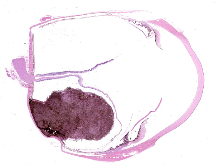
The vitreous was only partially present between a pigmented chorioidal mass and the completely ablated, cup shaped retina, that was only attached at the papilla nervi optici and focally attached to the posterior side of the lens.
Within the chorioidea, partly covered by the ablated retina, there was a nodular round to oval shaped endophytic protruding soft mass (about 10 mm in diameter) located between the pars ciliaris of the ciliary body and the papilla nervi optici with a homogenously black colored cut surface.
Laboratory Results:
- Urea: 1.499 mmol/l
- Creatinin: 12906.692 µmol/l
- Urea/Creatinin: 16
- Alanin-amino-transferase (ALT): 500.1 nkat/l
- Aspartat-amino-transferase (AST): 316.73 nkat/l
- Alkaline phosphatase (ALKP): 316.73 nkat/l
- Thyroxin: 25 nmol/l
- Serum chemistry profile values were within normal limits.
Microscopic Description:
The conjunctiva bulbi, cornea, anterior eye chamber, irido-corneal angle, the lens and the nervus opticus / optic cup do not show any histological changes.
Limbus: The limbal cornea shows a mild infiltration by pigmented cells (melanosis oculi).
Iris: There is moderate mydriasis, the facies posterior contains multifocal single cysts.
Ciliary body: The outer non-pigmented epithelium reveals a mild to focally moderate atrophy.
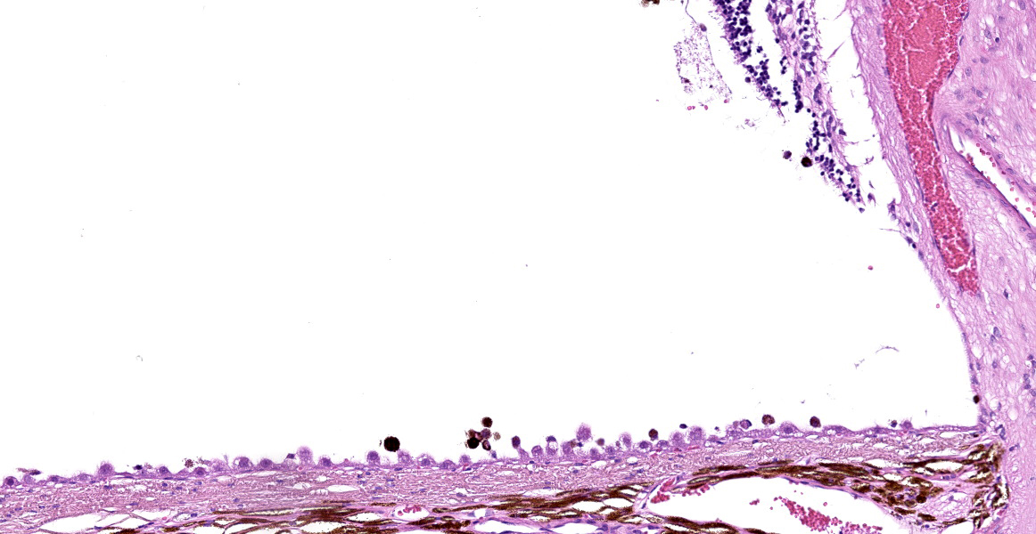
Vitreous body: There is focal evidence of scattered plump melanin containing cells (questionable RPE).
Retina: A total retinal ablation with moderate to severe atrophy of all retinal layers (neuronal - inner/outer granular-and photoreceptor layer) is present.
Retinal pigmentary epithelium (RPE): There is focally mild hypertrophy.
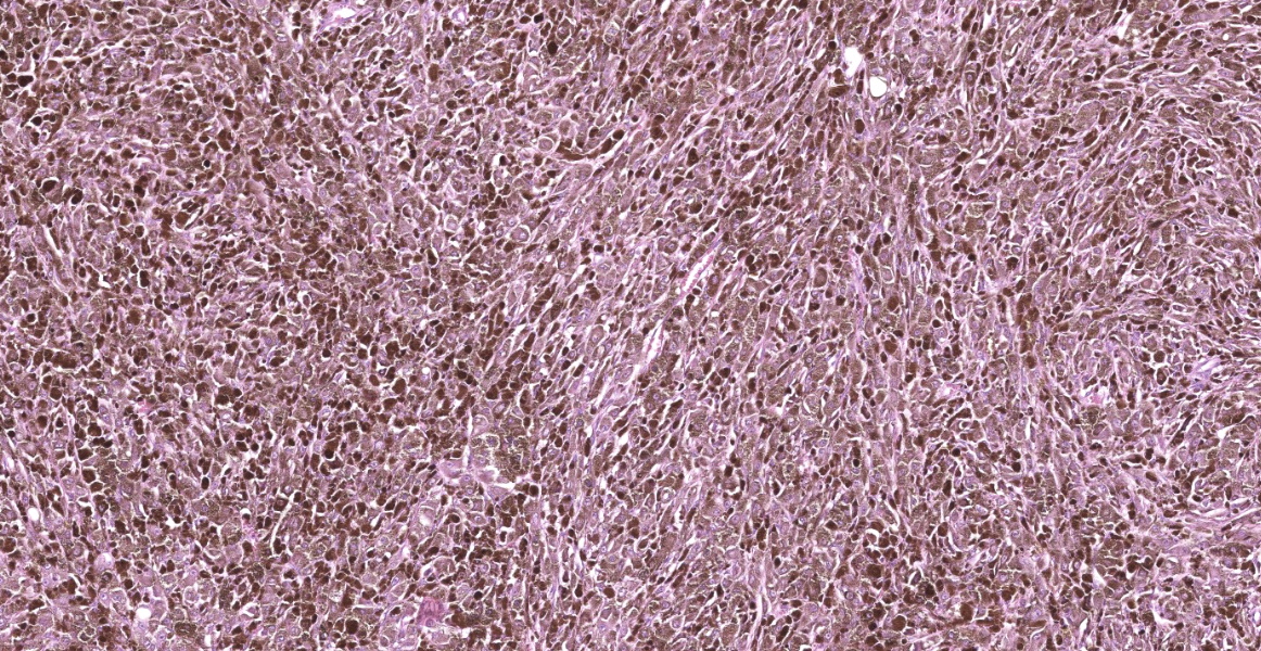
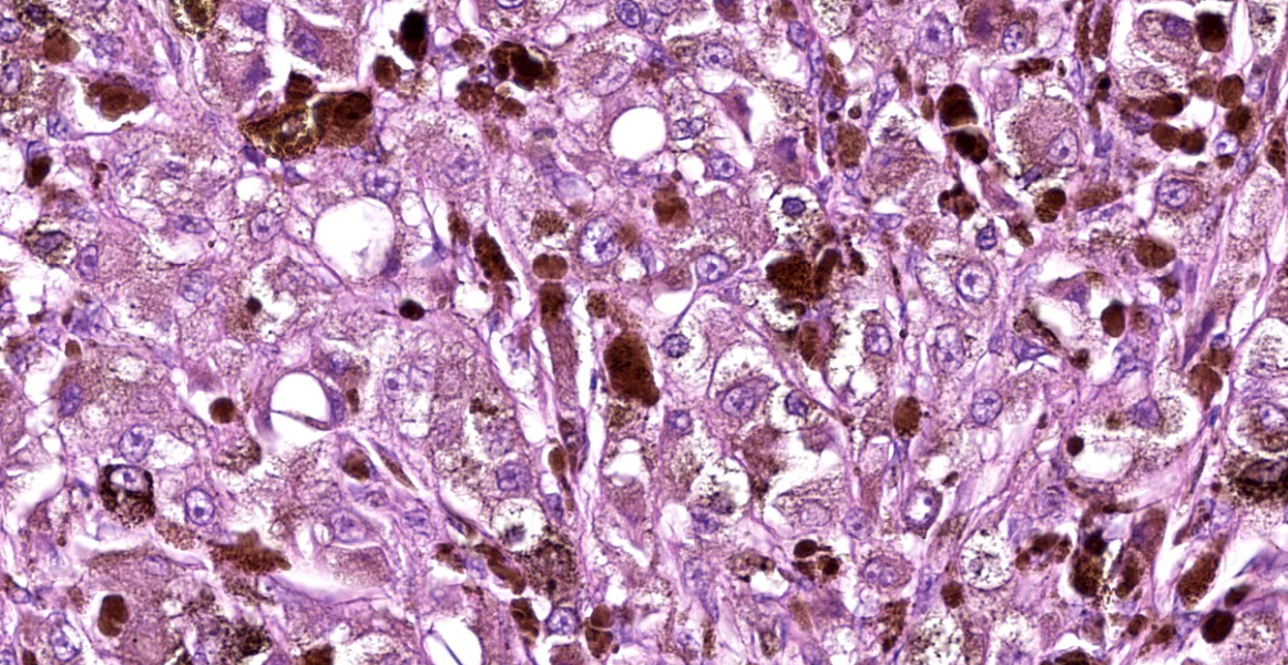
Chorioid: Located between ciliary body and papilla nervi optici, within the chorioid, there is a round to oval shaped, about 1 cm in diameter large chorioidal mass with focally infiltrative growth, partly covered by a thin layer of RPE and regular chorioidal pigmented cells (cream brown pigment). The cells are moderately densely packed, well demarcated and demonstrate solid to nodular growth pattern. The moderately pleomorphic cell population appears disordered, divided by a fine fibrovascular stroma. The cells are plump, round to weakly elongated with a diameter of about 25 µm, with eosinophilic cytoplasm with a variable content of cytoplasmatic granular, partly clumped brownish material (melanin granules) and often indistinct cell borders. Where discernible, the nuclei are about 10 µm in diameter and contain little condensed chromatin and one central nucleolus. Anisocytosis and anisokaryosis is high. Only few binucleated cells are visible, the bleached specimens show 0-1 mitotic figures per high power field. Focally, in the periphery of the cell proliferate there is a small area of osseous metaplasia.
Sclera: Opposing the tumor, the sclera is focal mildly thinned. There is no evidence of the neoplastic cells infiltrating the sclera.
Contributor’s Morphologic Diagnoses:
- Chorioid: endophytic choroidal plump cellular melanoma
- Ciliary body: atrophy of outer non-pigmented epithelium, mild to focally moderate
- Retina: total retinal ablation and atrophy, moderate to severe
- RPE: hypertrophy, focal mild
- Iris: iridal mydriasis with focal cyst formation (facies posterior), focal, moderate
- Melanosis oculi, focal, mild
Contributor’s Comment:
The most common melanocytic tumor in the eye of the cat is the feline diffuse iris melanoma.2 In the present case the neoplastic cell proliferate was not involving the iridociliary body and it was only focal, protruding into the vitreous. Additionally, iridociliary epithelial tumor can also be pigmented, but have been ruled out in this case due to same reasons as mentioned above.2
According to Parul Singh and Abhishek Singh`s Paper in 2012, choroidal melanoma is the most common malignant primary neoplasia in humans with most often metastasis in liver, frequent the lung and bone.6 Whereas canine choroidal melanomas are common, being usually benign and never metastasize2, a choroidal melanoma has been reported in a cat the first time in 2011.5 In the current case, no X-ray was performed to check for metastasis and no history of further locations with potential cell proliferates is known. The large cell proliferate led to ablation and atrophy of the retina with consecutive atrophy of the outer epithelium of the ciliary body and focal RPE hypertrophy, interpreted as secondary glaucomatous alterations.
Iridal cysts can be congenital or occur spontaneously, due to trauma or inflammatory processes.3 In the current case, it is seen as an incidental finding just as the mild melanosis oculi.
Contributing Institution:
Institute of Veterinary Pathology Zuerich (IVPZ)
www.vetpathology.uzh.ch
JPC Diagnosis:
- Eye: Choroidal melanocytoma.
- Eye: Iridal cysts.
JPC Comment:
While iris melanoma is the most common intraocular tumor of cats, this case is an example of melanoma originating from extra-iridal structures, which is uncommon to rare in cats.1,4 Only four cases of choroidal melanocytic neoplasms have been reported in the English literature. In two cases, neoplastic melanocytes infiltrated into the adjacent iris, and in a third case, there was a high mitotic rate (24 per 10 high power fields) with significant anisocytosis and anisokaryosis.1 These three cases were diagnosed as choroidal melanoma.1 The remaining case was diagnosed as a melanocytoma.5
Melanocytic neoplasms more commonly arise from the iris and are termed feline diffuse iris melanoma (FDIM).4 These neoplasms start as iris melanosis, characterized as a non-neoplastic focus of proliferative and dysplastic melanocytes up to 3 layers thick.4 Iris melanosis may remain static or rapidly progress to FDIM with infiltration of the subjacent iris stroma.4 Early FDIM and iris melanosis are grossly indistinguishable, but as FDIM progresses, the iris becomes thickened and the pupil may be irregularly shaped and less mobile.4 Features of malignancy for FDIM include a high mitotic rate (not well established and cut-off varies between studies), high nuclear to cytoplasmic ratio, significant nuclear pleomorphism, and multinucleation.1,4 Survival for cats with FDIM is correlated with histologic invasion, with neoplasms confined to the iris or trabecular meshwork producing survival times similar to control cats, and neoplasms extending into the sclera associated with decreased survival time.4 While cellular morphology can vary significantly and include patterns such as spindle cell, anaplastic, and giant cell variants, there is no correlation between morphology and metastasis.4 Rates of FDIM metastasis vary from 19 to 63%, and the liver is the most common site of metastasis.4 While some aggressive FDIMs metastasize quickly, others may metastasize late in the disease, sometimes years after the initial diagnosis.4
References:
- Bourguet A, Piccicuto V, Donzel E, Carlus M, Chahory S. A case of primary choroidal malignant melanoma in a cat. Vet Ophthalm. 2015; 18(4): 345-349.
- Dubielzig RR. Tumors of the Eye. In: Meuten DJ, ed. Tumors in Domestic Animals. 5th Ames, IO: Wiley-Blackwell; 2017:892-911.
- Gemensky-Metzler AJ, Wilkie DA, Cook CS. The use of semiconductor diode laser for deflation and coagulation of anterior uveal cysts in dogs, cats and horses a report of 20 cases. Vet Ophthalmol. 2004;7(5):360-8.
- Kayes D, Blacklock B. Feline Uveal Melanoma Review: Our Current Understanding and Recent Research Advances. Vet Sci. 2022; 9(2): 46-59.
- Semin MO, Serra F, Mahe V, Deviers A, Regnier A, Raymond-Letron I. Choroidal melanocytoma in a cat. Vet Ophthalmol. 2011;14(3):205-8
- Singh P, Singh P. Choroidal melanoma.Oman J Ophthalmol. 2012; 5(1):3-9.