Signalment:  

An adult, female, German shepherd dog (Canis familiaris)A 6-year-old female German shepherd dog was presented for a posterior ataxia and a bilateral quadriceps amyotrophy. At neurological examination, the posterior patellar reflex was bilaterally increased and the posterior proprioceptive reflexes were decreased. The dog was euthanized for humane reasons and a complete necropsy examination was undertaken.


Gross Description:  

At necropsy, a nonencapsulated, grayish granular mass was observed within the 4th ventricle. Nodular and multifocal intradural dull gray lesion with a granular cut surface was also observed on the ventral face of the brain stem scattering along the spinal cord. The nodules were firm, and had expansive growth without evidence of infiltration of the brain and spinal cord parenchyma. They compressed and displaced the spinal cord. 


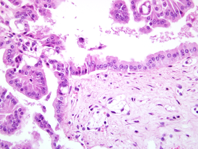
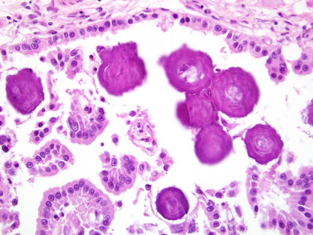
Histopathologic Description:

Spinal cord cross and sagittal sections: At histopathological examination, the ventral subarachnoidal space of the spinal cord and the nerve roots were severely infiltrated by a multinodular, poorly-demarcated, partially encapsulated mass with a moderate cellularity. The tumor was composed of papillary structures supported by a delicate fibrovascular stroma (Fig. 2-1). The neoplastic cells were cuboidal or columnar, measuring 15 to 20 μm with well-defined cytoplasmic borders. The cytoplasm was abundant and pale eosinophilic. The nucleus was round, basally located, euchromatic with coarse chromatin. The mitotic index was 3 to 4 mitoses per high power field and was dependent on examined area. Atypia cellular was moderate to strong: anisokaryosis and anisocytosis, nuclear gigantism, prominent nucleoli and multinucleated cells. No embolus was found. Numerous psammoma bodies and foci of mineralization were observed and focal perivascular accumulation of lymphoid cells was noted (Fig. 2-2). 


Morphologic Diagnosis:  

Spinal cord: Meningeal metastasis of choroid plexus carcinoma (meningeal carcinomatosis)


Lab Results:  

No significant bacterial pathogens were culture.

White blood cells count: 67 x 103 cells/mL (leucocytosis)
Red blood cells count: 2,3 x 10⁶ cells/mL (anemia)
Hematocrit : 18%
MCH: 63 g/L
Reticulocyte count : 50 %


Condition:  

Metastatic choroid plexus carcinoma


Contributor Comment:  

Choroid plexus tumors are usually rare and benign tumors. They have been described in man, cattle, horses, goats, cats, mice and dogs. The average age of affected dogs is 6 years and male dogs are up to three times more commonly affected than females, but there is no breed predisposition. In human pathology, choroid plexus carcinoma occurs mainly in children under 3 years of age.

The fourth ventricle is the most common site for these tumors in man and dog. In our case, a primary mass was detected microscopically in the choroid plexus of the fourth ventricle. No significant lesion was detected at post mortem examination so the possibility of metastasis of another tumoral process was ruled out. The spinal cord tumor was multiple without embolus so the hypothesis of a meningeal carcinomatosis due to diffusion of the neoplastic cells through cerebrospinal pathways is highly probable. Concerning immunohistochemistry, it is reported that Pankeratin and CK AE1 positivity is observed in choroid plexus carcinomas with marked cell anaplasia, whereas CK AE3 is expressed by well-differentiated neoplastic cells.1 Vimentin positivity is observed in a large number of neoplastic cells. EMA and S-100 give negative results in all cases of choroid plexus carcinoma.


JPC Diagnosis:  

Meninges, spinal nerve root: Metastatic choroid plexus carcinoma, German shepherd dog (Canis familiaris), canine.


Conference Comment:  

While some have proposed including a category of choroid plexus papilloma with atypia,4 two major forms of choroid plexus tumors are generally recognized: choroid plexus carcinoma and papilloma. Any form of anaplasia and/or metastasis, including metastasis of well-differentiated tumors within the ventricular system and along the neuraxis, is sufficient for a diagnosis of choroid plexus carcinoma.3 Anaplastic features include nuclear atypia, loss of papillary architecture with transition to patternless cellular sheets, an increased mitotic index, and necrosis.2,3

Papillary ependymomas can be included in the differential diagnosis for choroid plexus tumors. On H&E, ependymomas contain pseudorosettes, occasionally true rosettes with cilia, and have a glial rather than a fibrovascular core.2,3 Immunohistochemistry may be necessary to differentiate the two.

Table adapted from Ribas et al.4 and Koestner et al.2
Immunohistochemical stainEpendymoma (Papillary)Choroid plexus tumor
CytokeratinPositivePositive
GFAP (glial fibrillary acidic protein)PositiveUsually negative, but rarely positive


By immunohistochemistry performed at the AFIP, neoplastic epithelial cells in this tumor had positive cytoplasmic immunoreactivity to cytokeratin and GFAP. While most reported cases of canine choroid plexus tumor are negative for GFAP, at least one case in the literature was positive for GFAP.1 GFAP positivity of some canine choroid plexus tumors is not surprising based on their histogenesis and the findings in human choroid plexus tumors. Human choroid plexus tumors are often positive for both epithelial markers and glial markers reflecting their hybrid nature. Ependymomas should be widely positive for GFAP and are generally negative for cytokeratin.5


References:

1. Cantile C, Campani D, Menicagli M, Arispici M: Pathological and immunohistochemical studies of choroid plexus carcinoma of the dog. J Comp Pathol 126:183-93, 2002
2. Koestner A, Higgins RJ: Tumors of the nervous system. In: Tumors in Domestic Animals, ed. Meuten DJ, 4th ed., pp. 709-712. Blackwell Publishing, Ames, IA, 2002
3. Koestner A, Bilzer T, Fatzer R, Schulman FY, Summers BA, Van Winkle TJ: Tumors of neuroepithelial tissue. In: WHO International Histological Classification of Tumors of the Nervous System of Domestic Animals. 2nd series, vol. V, pp. 17-24, Armed Forces Institute of Pathology and American Registry of Pathology, Washington, DC, 1999
4. Ribas JL, Mena H, Braund KG, Sesterhenn IA, Toivio-Kinnucan M: A histologic and immunocytochemical study of choroid plexus tumors of the dog. Vet Pathol 26:55-64, 1989
5. Summers BA, Cummings JF, de Lahunta A: Tumors of the central nervous system. In: Veterinary Neuropathology. pp. 351-401, Mosby, St. Louis, Missouri, 1995

A virtual slide is not available for this case.


(Fig 2-1) Meninge


(Fig 2-1) Meninges


Back | VP Home | Contact Us |