Joint Pathology Center
Veterinary Pathology Services
Wednesday Slide Conference
2018-2019
Conference 20
20 March, 2019
CASE I: D12-49 (JPC 4041970).
Signalment: 8-year-old, male, castrated Labrador retriever (Canis lupus familiaris)
History: A cutaneous mass adjacent to the prepuce was surgically removed with wide margins and submitted for histologic evaluation. Following excision, the mass recurred and continued to grow despite treatment with prednisone and vinblastine. Therapy was switched to Palladia and resulted in partial remission for one month, at which point the neoplasm began to increase in size again. Euthanasia was elected due to poor prognosis and failed response to therapy.
Gross Pathology: None available.
Laboratory results: None available
Microscopic Description:
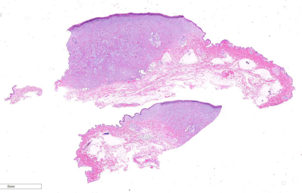
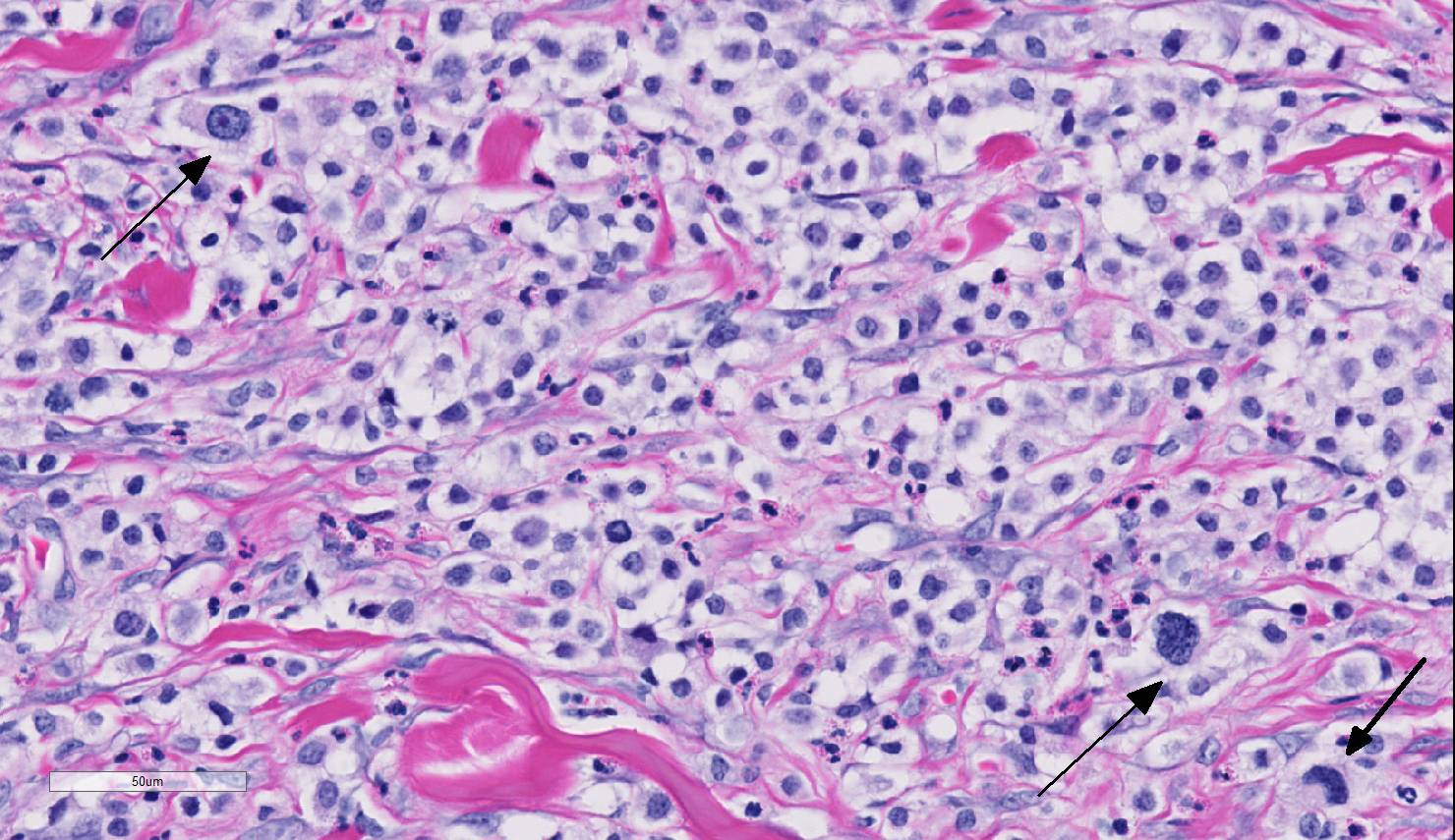
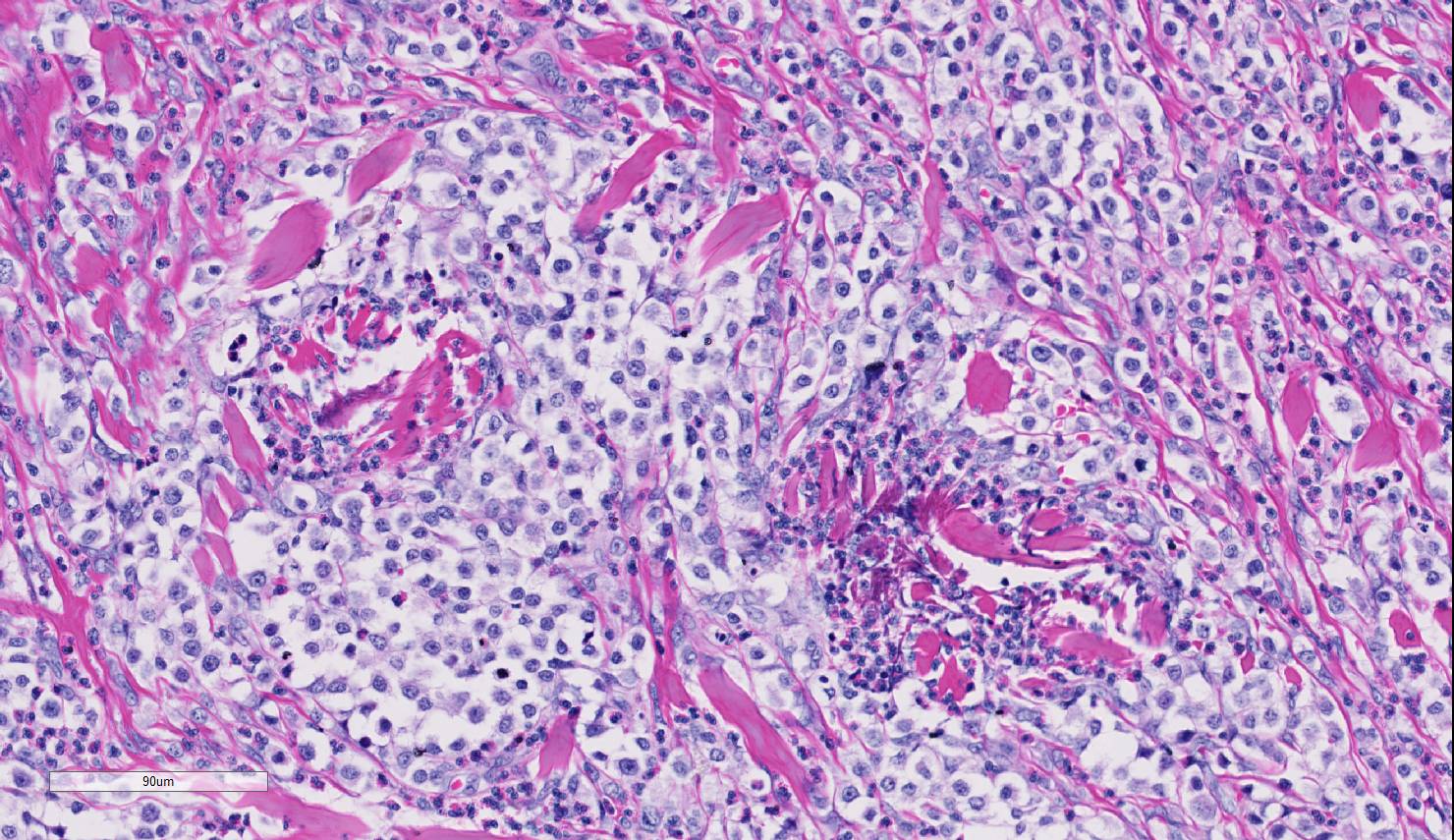
Expanding the dermis and elevating the overlying ulcerated epidermis is a well circumscribed, unencapsulated neoplasm consisting of numerous mast cells with marked anisocytosis and anisokaryosis, karyomegaly, bizarre nuclei, no apparent granules, 1-2 irregularly round nuclei, and a mitotic index of 5-6 per high power field, with bizarre mitotic figures. These cells are admixed with high numbers of eosinophils and multifocal collagenolysis (flame figures). The margins appear complete.
Contributor’s Morphologic Diagnoses:
Haired skin: Mast cell tumor, high grade.
Contributor’s Comment: Mast cell tumors account for 15-20% of skin tumors in dogs, and are the most frequent malignant tumor of the skin. In 2011, Kiupel et al. proposed a two-tier histologic grading system to more accurately predict the biological behavior of canine cutaneous mast cell tumors.2 This two-tier approach replaced the commonly used three-tier Patnaik system, and has since been shown to provide useful prognostic information with increased consistency of grading among veterinary pathologists.1,6.
This particular neoplasm has a very high mitotic rate with marked atypia of neoplastic cells, and serves as a prime example of a high grade neoplasm. Clinical follow-up on this patient revealed recurrence of the neoplasm at the site of excision and failure of response to treatment.
While not present in this case, it was recently reported that canine cutaneous mast cell tumors can exhibit epitheliotropism, and should be included as a differential for cutaneous round cell neoplasms with epitheliotropic behavior.4
Contributing Institution:
Tri-Service Research Laboratory
JBSA Ft. Sam Houston, TX
JPC Diagnosis: Haired skin: Mast cell tumor, high grade.
JPC Comment: In 2011, a 28-pathologist, 16-institution study on histologic grading of cutaneous mast cell tumors was published which has significantly increased concordance between pathologists in the grading and prognosis of canine mast cell tumors.2 This system is currently in use at the Joint Pathology Center and in most diagnostic laboratories. The study replaced previous 3-tier studies by Bostock (1973) and Patnaik (1984), both 3-tier studies in which interobserver variation resulted in an skewing of grading toward intermediate (grade 2) grades.2
The current two-tier system classifies cutaneous mast cell tumors into low- versus high-grade. The diagnosis of high grade mast cell tumors is based on the presence of ANY ONE of the following criteria: seven mitotic figures per ten 2.37mm2 fields, 3 multinucleated cells or cells with bizarre nuclei per ten 2.37mm2 fields, or karyomegaly (with nuclei of at least ten per cent of neoplastic cells varying by at least two fold. In this particular case, the participants were in agreement with an elevated mitotic count as well as the presence of an increased number of cells with bizarre nuclei.2 The count of mitotic figures should be performed in the areas of the slide with the highest frequency of mitotic figures.2
The kit protein is important in the proliferation, differentiation, migration and survival of mast cells. A variety of c-kit mutations have been identified in mast cell tumors, with the most common mutation (in exon 11) being associated with a significantly worse prognosis.3 In a study, 19/49 dogs with cutaneous mast cell tumors possessing a c-Kit mutation on exon 11 died within a year due to MCT-associated disease. Mutations on chromosome 8 and 9 compose less than 5% of overall mutations each, but have no impact on prognosis. PCR testing on this neoplasm was performed at the Michigan State Diagnostic Lab and was positive an activation duplication mutation in exon 11 of c-Kit, but was negative for exon 8 mutations.3
While mast cell tumors may arise in any organ, the skin is the most common site for mast cell tumor development in the dog. Another classification is important in evaluating cutaneous mast cells – that of a cutaneous versus subcutaneous location. Under previous grading systems, tumor depth was considered an adverse prognostic factor. Thompson et al. in 20115 evaluated followup data for 206 subcutaneous mast cell tumors (those restricted to the subcutaneous fat) and found that 6-month, 1-, 2- and 5-year survival times were 95%, 93%, 92%, and 86% respectively, indicating a significantly better prognosis for this subset of tumors. Only 4% exhibited metastasis, and 6% had local recurrence, although 56% of cases had incomplete tumor margins.5
References:
- Giantin M, Vascellari M, Morello MM, et al. c-KIT messenger RNA and protein expression and mutation in canine cutaneous mast cell tumors: correlations with post-surgical prognosis. J Vet Diagn Invest. 2012;24(1):116-126.
- Kiupel M, Webster JD, Bailey KL, et al. Proposal of a 2-Tier Histologic Grading System for Canine Cutaneous Mast Cell Tumors to More Accurately Predict Biological Behavior. Vet Pathol. 2011;48(1):147-155.
- Kiupel M. Mast cell tumors. In Meuten DJ, ed. Tumors in Domestic Animals, 5th ed. Ames, IA, Wiley Blackwell, pp. 177-177-193.
- Oliveira FN, Elliott JW, Lewis BC, et al. Cutaneous Mast Cell Tumor With Epitheliotropism in 3 Dogs. Vet Pathol. 2013;50(2):234-237.
- Thompson JJ, Pearl DL, Yager JA, Best SJ, Coomber BL Foster RA. Canine subcutaneous mast cell tumor: characterization and prognostic indices. Vet Pathol 2011; 48(1):156-168.
- Vascellari M, Giantin M, Capello K, et al. Expression of Ki67, BCL-2, and COX-2 in Canine Cutaneous Mast Cell Tumors: Association With Grading and Prognosis. Vet Pathol. 2013;50(1):110-121.