Signalment:
3-year-old female mixed breed pig (
Sus scrofa domestics).The animal was slaughtered in an officially inspected abattoir in Catalonia (Spain).
Gross Description:
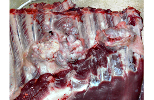
Two prominent exophytic, multinodular, cauliflower shaped masses of 4 and 5 cm in diameter were noticed in the inner side of the thoracic cavity arising from the rib surface. The masses had a smooth surface and were attached with a broad sessile base to the costal body of the ribs. The masses were solid, had a hard consistency and at cross section had bluish to white areas. During the inspection, no other abnormalities were observed in the carcass or in the internal organs of the animal.
Histopathologic Description:
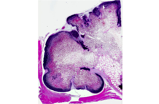
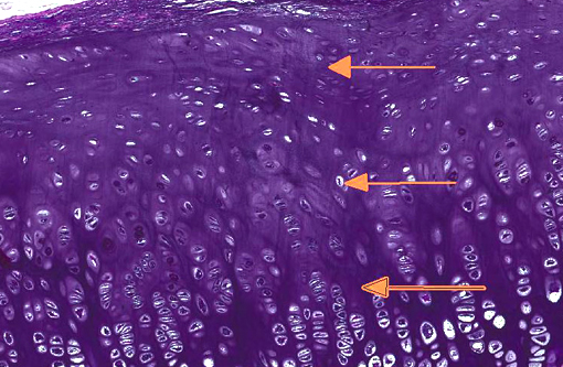
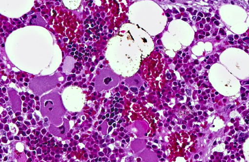
Rib, transverse section: There is an exophytic, broad based, multinodular, well demarcated, expansive, and non-encapsulated neoplastic proliferation arising from the rib poriosteum. Every nodule is lined by fibrovascular stroma of variable thickness (perichondria). Neoplastic cells are well-differentiated chondrocytes enmeshed within an abundant hyalinized amphophilic extracellular matrix. They are arranged from the periphery to the center in well-defined layers mimicking growth plate endochondral ossification. The outer layer is made up of multiple small groups of chondrocytes with high nucleus/cytoplasm ratio (resting layer). Secondly, there is a thicker layer constituted by larger chondrocytes forming columns (proliferative layer), and a deeper layer where chondrocytes undergo hypertrophy (hypertrophic layer). Underneath the hypertrophic layer, the cartilage matrix is replaced by hyaline, eosinophilic osteoid matrix (calcification layer) and subsequently, there is loss and replacement of the chondrocytes by osteocytes. This is partially lined by osteoclasts and rows of plump osteoblasts. In the center of the nodule, several septa of mature trabecular bone are seen. Amongst the septa, bone marrow with a heterogeneous population of hematopoietic precursors and scattered adipocytes are present.
Morphologic Diagnosis:
Rib osteochondroma.
Condition:
Osteochrondroma
Contributor Comment:
An osteochondroma is a benign, cartilage-capped tumor arising from the surface of bones formed by endochondral ossification. Osteochondromas may occur in two forms: solitary or multiple.(2,5) This condition is usually recognized as an incidental finding during routine controls, radiographic examination or at necropsy. Occasionally, clinical signs might occur and are due to compression or distortion of adjacent structures. Osteochondromatosis is infrequently reported in humans, horses, dogs, a macaque and cats.(2,3,6) In the present case, considered differential diagnoses included chondroma and chondrosarcoma. Histologically, chondroma consists of irregular lobules of hyaline cartilage which may also show foci of endochondral ossification and mineralization.(7,8) However, chondroma misses the growth plate-like organization of the cartilaginous matrix that characterizes osteochondroma. Malignant transformation of osteochondroma to chondrosarcoma has occasionally been described in older dogs and humans. Histologically osteochondroma can be very difficult to differentiate from low grade chondrosarcoma, which may show few indications of malignancy and may closely resemble benign tumors of cartilage.Â
JPC Diagnosis:
Rib: Osteochondromas (multiple cartilaginous exostoses).Â
Conference Comment:
Osteochondroma are typically continuous with the marrow cavity of the underlying bone, a feature which is helpful in differentiation from other proliferative or neoplastic bone lesions.(8) This lesion is relatively uncommon in swine, however it occurs fairly frequently in dogs and horses, where it is inherited in an autosomal dominant pattern and typically arises from scapula, ribs, vertebrae, and pelvis of young animals.(1) Although the underlying genetic defect in these species is unknown, it likely involves the perichondrial ring. Since enlargement ceases at the time of physeal closure, and the cartilage cap is eventually replaced by bone in an orderly fashion, there is some debate regarding its classification; many do not consider osteochondroma to be a true neoplasm, but rather a skeletal dysplasia.(8) As a result, the terminology associated with osteochondromas can be somewhat confusing. Osteochondromatosis is also known as multiple osteochondromas, multiple hereditary exostoses, multiple cartilaginous exostoses, multiple osteochondromatosis, diaphyseal aclasis, and hereditary chondrodysplasia.(4)
In contrast, feline osteochondromatosis occurs in older animals and primarily affects intramembranous flat bones, while long bones are seldom affected. The lesion does not generally communicate with the marrow cavity of the underlying bone. Cells may appear somewhat atypical and pleomorphic, and growth tends to be progressive, with the potential for malignant transformation and metastasis. Thus feline osteochondromatosis is more consistent with a true neoplasm and has a more guarded prognosis. Inheritance does not appear to play a role in the pathogenesis; instead, viral particles resembling feline leukemia virus (FeLV) have been demonstrated within the proliferating cells of the hyaline cartilage cap in these tumors.(1)
References:
1. De Brot S, Grau-Roma L, Vidal E, Segales J. Occurence of osteochondromatosis (multiple cartilaginous exostoses) in a domestic pig (Sus scrofa domesticus). J Vet Diagn Invest. 2013;25(5):599-602.
2. Franch J, Font J, Ramis A, et al. Multiple cartilaginous exostosis in a Golden Retriever cross-bred puppy. Clinical, radiographic and backscattered scanning microscopy findings. Vet Comp Orthop Traumatol. 2005;18:189-193.
3. Matthews KA, Strait K, Connor-Stroud F, Courtney CL. Osteochondromatosis in a Rhesus Macaque (Macaca mulatta). Comp Med. 2012;62:149-152.
4. Ranade SA, Pacchiana PD. What is your diagnosis? Osteochondromatosis. J Am Vet Med Assoc. 2011;238(10):1243-1244.
5. Romeo S, Hogendoorn PC, Dei Tos AP. Benign cartilaginous tumors of bone: from morphology to somatic and germ-line genetics. Adv Anat Pathol. 2009;16:307-315.
6. Saglik Y, Altay M, Unal VS, et al. Manifestations and management of osteochondromas: a retrospective an+�-�lisis of 382 patients. Acta Orthop Belg. 2006;72:748-755.
7. Thompson KG, Pool RR. Tumors of bone. In: Meuten DJ, ed. Tumors of Domestic Animals. 4th ed. Ames, Iowa: Iowa State Press; 2002:245-317.
8. Thompson KG. Bones and joints. In: Maxie MG, ed. Jubb, Kennedy, and Palmers Pathology of Domestic Animals. 5th ed. Vol. 1. St. Louis, MO: Elsevier Limited; 2007:118-124.