Signalment:
6-year-old Angus cow (
Bos taurus).The herd has been out at pasture with no significant abnormalities described. The owner noticed
excessive hair loss on this animal several days prior to its acute death.
Gross Description:
A 1050 pound adult female cow is presented for necropsy and most of the carcass is noted to be
alopecic. In multifocal to coalescing foci the cardiac muscle is tan to pale yellow and slightly raised on cut section.
Histopathologic Description:
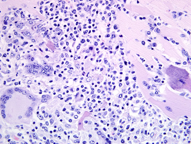
In multifocal to coalescing foci there is extensive replacement of the epicardium and
myocardium by predominately a granulomatous and lymphocytic infiltrate with fewer eosinophils and plasma cells.
Multinucleated giant cells containing up to 30 nuclei (both foreign body type and Langhans type) are common (
fig. 4-1).
Degenerative changes in adjacent myofibers include hypereosinophilia, loss of cytoplasmic detail (cross striations)
and mineralization. A similar type of inflammatory infiltrate was found histologically in the kidney, liver, adrenal
gland, and skin. PAS, Grams stain, and Acid-Fast staining failed to illustrate etiologic agents.
Morphologic Diagnosis:
Myocarditis, granulomatous and lymphocytic, multifocal to coalescing,
chronic, severe.
Lab Results:
Only postmortem contaminants were isolated via bacterial culture of the myocardium.
Condition:
Hairy vetch toxicosis
Contributor Comment:
The multiorgan distribution of a granulomatous infiltrate described in this case and the
failure to identify causative etiologic agents is consistent with hairy vetch (
Vicia villosa) toxicosis. Hairy vetch is a
forage legume and can be commonly found in pastures in the United States and other temperate regions of the world.
It is widely cultivated and used as pasturage, harvested as hay and silage, and utilized as a cover crop although it has
been associated with a systemic granulomatous disease in some cattle. The specific etiologic factors and pathogenic
mechanisms involved in the development of this syndrome remain unknown. It is unusual that hairy vetch is
commonly ingested by cattle without the development of clinical signs and it is difficult to routinely induce the
disease experimentally. It has been proposed that a plant constituent (lectins) induces a type IV, or cell mediated
hypersensitivity reaction or potentially directly activates T lymphocytes initiating the systemic granulomatous
reaction described with the syndrome. Furthermore, the low incidence of disease and disease occurrence when
availability of vetch is limited suggest duration of exposure or repeated exposure to the plant help support a
hypersensitivity theory. As the syndrome mainly occurs in Holstein and Angus cattle a genetic predisposition has
also been suggested.(2,3)
Cattle that develop clinical disease usually have been grazing a pasture that contains a mixture of hairy vetch and
small grains, including rye, wheat, and oats and outbreaks typically occur during the season of maximal vetch
growth.(2) Additionally, development of the disease occurs after grazing an affected pasture for at least 2-6 weeks.
Vetch associated disease is typically more severe in adult cattle greater than 3 years old.
The clinical syndrome associated with Vetch toxicosis varies and three different clinical manifestations have been
described. One syndrome involves acute neurological signs and death consistent with cyanogenic glycosides
contained in the seeds. A second syndrome involves subcutaneous swellings of the head, neck, and body with ulcers
of the oral mucous membranes, purulent nasal discharge, rales, cough and congestion. The third syndrome involves
the systemic granulomatous response and presents clinically as a dermatitis and conjunctivitis with diarrhea and
weight loss.(3) With the systemic granulomatous syndrome gross lesions include a multifocal to coalescing, soft,
and gray to yellow infiltrate disrupting the normal architecture of the involved tissues (liver, kidney, spleen, heart,
adrenal gland, skin). Histologically, the salient feature is a multifocal to coalescing granulomatous infiltrate
disrupting the affected tissue architecture. The infiltrate is typically characterized by epitheloid macrophages,
multinucleated giant cells, lymphocytes, plasma cells and typically eosinophils.
JPC Diagnosis:
1. Heart: Myocarditis, granulomatous and eosinophilic, multifocal to coalescing, severe, with
myocyte degeneration and necrosis.
2. Heart, myocardium: Sarcocysts, few.
Conference Comment:
The contributor provided an excellent overview of hairy vetch toxicosis. As the
contributor noted, the pathogenesis of hairy vetch toxicosis remains somewhat unclear, and cattle exposed to the
plant do not consistently develop disease. Therefore, hairy vetch toxicosis remains a diagnosis of exclusion.
Conference participants discussed the importance of ruling out other causes of granulomatous inflammation, even
when affected cattle are known to be exposed to hairy vetch. Special stains for bacterial and fungal etiologies were
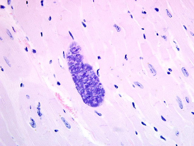
performed by the contributor, and repeated at AFIP, with no causative organisms seen. Some slides contained low
numbers of sarcocysts that were not associated with inflammation, and were considered incidental (
fig. 4-2).
Other compounds that may induce the production of lesions indistinguishable from vetch toxicosis include diureido-
isobutane (DUIB) and citrus pulp.(1) In horses, vetch toxicosis causes lesions similar to those in cattle, with
the notable exceptions being the relative lack of eosinophils in the infiltrate and the lack of heart involvement.(1)
References:
1. Ginn PE, Mansell JEKL, Rakich PM: Skin and appendages.Â
In: Jubb, Kennedy, and Palmers Pathology of
Domestic Animals, ed. Maxie MG, 5th ed., vol. 1, pp. 686-702. Elsevier Saunders, Philadelphia, PA, 2007
2. Johnson B, Moore J, Woods LW, Galey FD: Systemic granulomatous disease in cattle in California associated
with grazing hairy vetch (
Vicia villosa). J Vet Diagn Invest 4:360-362, 1992
3. Panciera RJ, Mosier DA, Ritchey JW: Hairy vetch (
Vicia villosa Roth) poisoning in cattle: Update and
experimental induction of disease: J Vet Diagn Invest 4:318-325, 1992