Signalment:  

7-month-old, female, neutered, German Shepherd Dog (Canis familiaris).This dog was singly kenneled in a training center which had 60 other dogs. This dog was found weak and ataxic, with vomiting and diarrhea and was euthanized. It was reported to be eating and drinking and to have had normal feces the day before. Ovariohysterectomy had been performed on this animal 10 days prior to euthanasia. A transitory diarrhea that resolved with oral metronidazole was present for a few days after surgery but no other complications were reported. All other dogs at the facility remained clinically normal.


Gross Description:  

Approximately 150 ml of sero-sanguineous fluid and large fibrinous clots were present in the thoracic cavity. There were numerous fibrinous adhesions between the visceral and parietal pleura and petechial hemorrhages were present in the intercostal muscles. The ventral 50-80% of all lung lobes was firm, black and depressed. Randomly scattered within these dark areas, were numerous white to pale pink, 1-4mm diameter irregular foci. The pericardial sac was thickened and edematous, with fibrinous adhesions to the visceral pleura. 


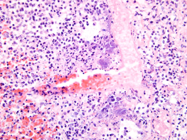
Histopathologic Description:

Most alveoli and bronchioles contain extravasated erythrocytes, eosinophilic proteinaceous fluid, fibrin and an inflammatory exudate of viable and necrotic neutrophils, with fewer macrophages and lymphocytes. There is extensive coagulative septal necrosis, with foci of complete parenchymal dissolution and replacement by necrotic cellular debris. In some sections focally extensive hemorrhage disrupts the normal architecture of the lung. Blood vessel necrosis and fibrin thrombosis are prominent in these areas. Colonies of short rod-shaped bacteria are present in many bronchioles and scattered throughout the necrotic parenchyma (fig. 4-1). There are scattered clumps of amorphous, basophilic material (consistent with mineral) in alveolar spaces. There are extensive subpleural hemorrhages. 

In the tracheobronchial lymph nodes there was lymphocellular necrosis, hemorrhage and medullary hemosiderosis. Widespread, acute centrilobular hepatic necrosis was the only other significant finding.


Morphologic Diagnosis:  

Lung: Pneumonia, necrotizing and fibrinohemorrhagic, acute, diffuse, severe with thrombosis and rod-shaped bacteria, etiology consistent with necrotoxigenic Escherichia coli.


Lab Results:  

A pure culture of hemolytic Escherichia coli was isolated from the lung. Serotyping classified the isolate as O4:H5 or O4:H56 and it tested positive for cytotoxic necrotizing factor 1 (CNF1). Samples of lung were negative for canine influenza by reverse-transcription PCR (RT-PCR).


Condition:  

E. coli pneumonia


Contributor Comment:  

Escherichia coli is the predominant facultatively anaerobic enteric bacterium of most vertebrates and is frequently isolated from diagnostic specimens. Most strains are either commensals or opportunistic pathogens of immunocompromised individuals, but other strains are well recognized pathogens. The pathogenic strains are historically classified into enteropathogenic, enterotoxigenic, enteroinvasive, enterohemorrhagic, necrotoxigenic and enteroaggregative strains according to the O (lipopolysaccharide) and H (flagellum) serotype, and are further characterized by their production of virulence factors5. Cytotoxic necrotizing factor 1 (CNF1) is consistently produced by necrotoxigenic E. coli and some isolates also produce CNF2 and alpha hemolysin. Necrotoxigenic strains are an important cause of extraintestinal disease such as urinary tract infections, pyometra, meningitis, septicemia and pneumonia in humans and mammals5. 

Hemorrhagic and necrotizing pneumonia caused by necrotoxigenic E. coli has recently been described in dogs1,4. In common with the present case, affected dogs have been young (<1year), the clinical illness is usually less than 24 hours and immunodeficiency or concurrent illness were not identified. In our case, parainfluenza virus and adenovirus testing were not performed and while no inclusion bodies were identified, concomitant infection with these agents cannot be excluded. E. coli of both O4 and O6 serotypes have been reported to cause these lesions, which irrespective of serotype were positive for CNF1. 

The main differential diagnoses for hemorrhagic pneumonia in dogs are canine influenza and bacterial septicemias including streptococcal septicemia 2,3. Microscopically, canine influenza is characterized by a pneumonia that is more broncho-interstitial and suppurative; with less necrosis 2, but RT-PCR or virus isolation is best performed for definitive exclusion. Samples of lung from this case were negative for canine influenza by RT-PCR.

Little is known about the source and route of infection, means of transmission and pathogenesis of this disease in dogs. 


JPC Diagnosis:  

Lung: Pneumonia, necrohemorrhagic, neutrophilic and histiocytic, diffuse, severe, with fibrin, edema, and numerous bacilli, German Shepherd Dog (Canis familiaris), canine.


Conference Comment:  

Strains of E. coli are identified by the various antigens they express, primarily using the O and H antigens. 
    O antigens (somatic): Determines the serogroup, lipopolysaccharide molecule
   H antigens (flagellar): Determines the serotype
   K antigens (capsular): Made up of polysaccharides and proteins; may also be used for classification purposes
   Fimbrial or pili antigens: Important in adhesion and colonization of epithelium

Extraintestinal pathogenic E. coli have been associated with pyometra, mastitis, otitis, prostatitis, bacteremia, skin diseases, cholecystitis, and pneumonia. Strains producing the cytotoxic necrotizing factor (CNF) are referred to as necrotoxic E. coli.4 These strains produce either CNF1, identified in humans and domestic animals, or CNF2, identified only in ruminants.4,5 The genes that code for CNF-1 and alpha hemolysin are genetically linked and have a tendency to occur with O4 and O6 groups.1,4

The primary fimbrial antigen in extraintestinal pathogenic E. coli is the P fimbriae and is encoded by the pap (pilus-associated pyelonephritis) gene.1,4 The papG (fimbrial tip adhesion) and papA (major fimbrial subunit) alleles have also been associated with necrotoxic E. coli.1,4


References:

1. Breitschwerdt EB, DebRoy C, Mexas AM, Brown TT, Remick AK: Isolation of necrotoxigenic Escherichia coli from a dog with hemorrhagic pneumonia. J Am Vet Med Assoc 226:2016-2019, 2005
2. Crawford PC, Dubovi EJ, Castleman WL, Stephenson I, Gibbs EP, Chen L, Smith C, Hill RC, Ferro P, Pompey J, Bright RA, Medina MJ, Johnson CM, Olsen CW, Cox NJ, Klimov AI, Katz JM, Donis RO: Transmission of equine influenza virus to dogs. Science 310:482-485, 2005
3. Garnett NL, Eydelloth RS, Swindle MM, Vonderfecht SL, Strandberg JD, Luzarraga MB: Hemorrhagic streptococcal pneumonia in newly procured research dogs. J Am Vet Med Assoc 181:1371-1374, 1982
4. Handt LK, Stoffregen DA, Prescott JS, Pouch WJ, Ngai DT, Anderson CA, Gatto NT, DebRoy C, Fairbrother JM, Motzel SL, Klein HJ: Clinical and microbiologic characterization of hemorrhagic pneumonia due to extraintestinal pathogenic Escherichia coli in four young dogs. Comp Med 53:663-670, 2003
5. Garnett NL, Eydelloth RS, Swindle MM, Vonderfecht SL, Strandberg JD, Luzarraga MB: Hemorrhagic streptococcal pneumonia in newly procured research dogs. J Am Vet Med Assoc 181:1371-1374, 1982
6. Songer JG, Post KW: The genera Escherichia and Shigella. In: Veterinary Microbiology - Bacterial and Fungal Agents of Animal Disease, eds. Songer JG, Post KW, pp. 113-119. Elsevier Saunders, St. Louis, MO, 2005

A virtual slide is not available for this case.


fig. 4-1 Lung


Back | VP Home | Contact Us |