Signalment:
Gross Description:
Histopathologic Description:
Morphologic Diagnosis:
Condition:
Contributor Comment:
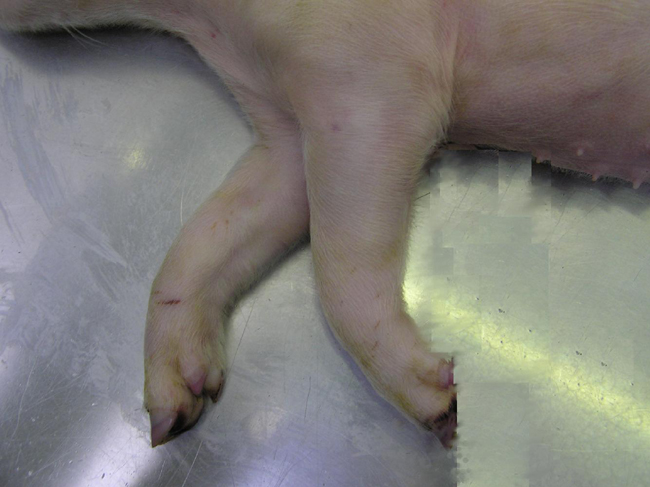
The radioulnar region is thickened and may be twice normal diameter. The overlying skin is hyperaemic. At necropsy there is enlargement of the soft periosseous tissues mostly at the cranial surface of the limbs from proximal end of the radius down to and surrounding the metacarpal bone distally. Fibrous tissue may invade and partially fuse with muscles 3.Â
Microscopically there are radiating osseous spicules associated with marked hyperplasia of periosteal osteoblasts. They are numerous, large and can appear as syncytium. Normal cortical bone is lamellar and forms concentric osseous plates consistent with rapid periosteal apposition. Osseous trabeculae of woven bone are orientated radially in relation to the medullary cavity3.Â
The pathogenesis of congenital hyperostosis is not known, however there are two suggested mechanisms reported in the piglet. These include 1) disruption of the growth of bone at the ossification groove of Ranvier or 2) local circulatory abnormality.Â
Dalton et al 3 proposed that radioulnar hyperostosis may be the result of an initial lesion situated at the anchor site of the periosteum to the epiphysis at the level of the perichondrial ossification groove of Ranvier. This true separation could be the cause of the fine supernumerary trabeculae of woven bone. The groove of Ranvier is an ossification groove (a component of the perichondrial ring supporting the zone of provisional calcification) that supplies chondrocytes to the physis for diametric growth of the foetal bone and also fibrous attach of the periosteum to the epiphysis.
A second study of piglets with hyperostosis, conducted by Roels et al 4 demonstrated distinct circular constrictions in the proximal antebrachial region of the median artery, in conjunction with the consistent finding of oedema within the connective tissue surrounding the thickened bone suggesting hypertension. In Affected piglets the initial segment of the median artery (ie proximal antebrachial region) showed distinct circular constrictions (not seen in controls) suggesting acute hypertension. There was extensive smooth muscle fibre proliferation, intimal fibrosis and fibrinoid necrosis of tunica media and less narrowing of lumen in small arteries and arterioles of upper dermis
JPC Diagnosis:
Conference Comment:
A similar hyperostotic condition has been reported in a single West Highland White Terrier dog, in which new bone formation involved the pelvis, scapulae, humeri, ulnae, femora, radii, and tibiae.5
The deeper, older portion of the examined cortex is formed of woven bone. This type of bone is formed in areas in which a support structure needs to be put in place quickly, such as in the developing fetus or at sites of fracture repair, inflammation, or neoplasia. It consists of collagen fibers within the bone matrix that are arranged in a haphazard interwoven fashion.5 These haphazard arrangements are usually replaced with the more structurally sound lamellar bone during skeletal maturation.5 In young rapidly growing animals, especially ruminants, a different type of lamellar bone is deposited along the surfaces of long bones. This type of bone is called laminar bone and consists of laminar arrays rather than the Haversian system seen more commonly in lamellar bone.5
References:
2. Dewey CE: Disease of the nervous and locomotor systems. In: Diseases of Swine, eds. Straw BE, Zimmerman JJ, DAllaire S, Taylor DJ, 9th ed., p. 88, Blackwell Publishing, Ames, IA, 2006
3. Doize B, Martineau GP: Congenital hyperostosis in piglets: a consequence of a disorganization of the perichondrial ossification groove of Ranvier. Can J Comp Med 48:414-419, 1984
4. Roels S, Simoens P, Ducatelle R: Localised arteriosclerotic changes in congenital hyperostosis in pigs. Vet Rec 139:446-447, 1996
5. Thompson K: Bones and joints. In: Jubb, Kennedy, and Palmers Pathology of Domestic Animals, ed. Maxie MG, 5th ed., vol. 1, pp. 6-7, 40-41, 106-110. Elsevier Limited, St. Louis, MO, 2007
6. Weisbrode SE: Bones and joints. In: Pathologic Basis of Veterinary Disease, eds. McGavin MD, Zachary JF, 4th ed., pp. 1066-1067. Elsevier, St. Louis, MO, 2007