Signalment:
Gross Description:
Histopathologic Description:
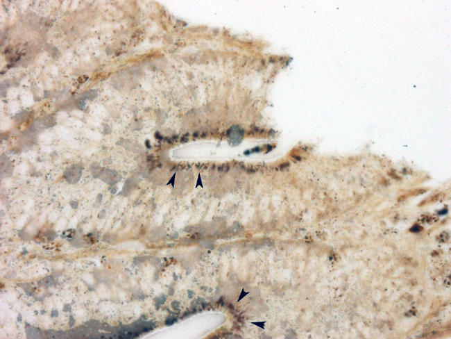
Steiner's silver technique shows numerous short, curved rods within the apical portions of glandular epithelial cells.
Morphologic Diagnosis:
Lab Results:
Condition:
Contributor Comment:
As an obligate, intracellular pathogen, pathogenesis of L intracellularis related disease requires active uptake by intestinal epithelial cells. Localization of lesions to the ileum may be related to uptake of organisms by epithelium associated with Peyer's patches. Organism are initially taken up in membrane-bound vesicles and later released into the cytoplasm where they multiply. Cell division is required for bacterial proliferation. The mechanism by which L intracellularis stimulates proliferation and dedifferentiation of ileal epithelial cells are poorly understood. Studies have shown, however, that the organism suppresses the inflammatory response by decreasing both B cell and T cell numbers, while macrophage numbers increase 4.Â
Gross lesions are characteristic of the various forms of the disease. Proliferative ileitis, also called intestinal adenomatosis, is characterized by ridge-like thickening of terminal portion of the ileum, occasionally extending cranially or caudally to involve the cecum and proximal spiral colon. The marked thickening can be observed from the serosal surface as accentuation of the normal reticular pattern of the ileum. Necrotic enteritis is characterized by coagulative necrosis of the adenomatous mucosa, likely the result of anaerobic bacterial proliferation. Chronic infection, ulceration and stricture may result in a lesion called regional ileitis, characterized by severe hypertrophy of the muscular layers of the ileum. Proliferative hemorrhagic enteropathy may occur when extensive necrosis and ulceration causes massive hemorrhage into the lumen of the ileum. Grossly, the typical adenomatosis lesion is overlain by clotted blood and fibrin. Animals affected by this form of the disease may die acutely from exanquination.
In all forms of the disease, the histologic features are similar. The characteristic morphology is that of marked hyperplasia of intestinal crypt epithelium with loss of goblet cells and minimal inflammation. Mitotic activity is high and glands become crowded, branched and dilated by accumulation of necrotic debris. Hyperplastic glands may protrude into the lymphoid follicles of the submucosa (a prominent feature in this case).
Differential diagnoses for diarrhea and weight loss in feeder pigs include swine dysentery and salmonellosis, both of which have distinct gross and histologic lesions centerd mainly on the cecum and colon. Porcine circovirus-2, the agent of postweaning multisystemic wasting disease PMWR), reportedly can cause similar histologic lesions in the absence of co-infection with L intracellularis2. That intestinal lesion is characterized by a necrotic, proliferative enteritis with marked replacement of Peyer's patches by histiocytes and multinucleate giant cells, which can also be a feature of PE. However, characterisitic botryoid cytoplasmic inclusions of PCV-2 infection should help differentiate the 2 diseases. No PCV-2 inclusions were seen in this case.
Although primarily a disease of pigs, L intracellularis can infect many species, most notably young horses, causing a similar proliferative enteropathy. The organism has also been investigated as an agent of inflammatory bowel disease in human beings 6.
JPC Diagnosis:
Conference Comment:
References:
2. Jensen TK, Vigre H, Svensmark B, Bille-Hansen V: Distinction between porcine circovirus type 2 enteritis and porcine proliferative enteropathy caused by Lawsonia intracellularis J Comp Path 135:176-182, 2006.
3. Lawson, GHK and Gebhart CJ: Proliferative enteropathy J. Comp. Pathol 122: 77- 100, 2000
4. MacIntyre N, Smith DGE, Shaw DJ, Thomson JR, Rhind SM: Immunopathogenesis of experimentally induced proliferative enteropathy in pigs. Vet Pathol 40:421-432, 2003
5. McOrist S, Gebhart CJ: Proliferative enteropathies. In: Diseases of Swine, eds. Straw BE, Zimmerman JJ, DAllaire S, Taylor DJ pp. 727-737. Blackwell Publishing, Ames, IA, 2006
6. Mickalski CW, Francesco Di Mola F, Kummel K, Wendt M, Koniger JS, Giese T, Giese NA, Friess H: Human inflammatory bowel disease does not associate with Lawsonia intracellularis infection. BMC Microbiology 6:81-88, 2006
7. Straw BE, Dewey CE, Wilson MR: Differential diagnosis of disease. In: Diseases of Swine, eds. Straw BE, Zimmerman JJ, DAllaire S, Taylor DJ pp. 727-737. Blackwell Publishing, Ames, IA, 2006