Signalment:
Adult, female crocodile (
Crocodylus niloticus).This crocodile was kept in a local zoo for over 2 years and was listless and anorexic for 5 weeks.
Gross Description:
All lung lobes contained multiple, 0.5-1.0 cm diameter, irregularly-shaped, well-defined, firm,
homogenous white to yellow nodules. Several firm, large nodules, 2.0-2.5 cm in diameter, white to yellowish in
color, surrounded by dark red foci, were found in some part of the lungs. Visceral surfaces in the lung and pleural
surfaces appeared dull and rough with a yellowish fibrous appearance. The liver and spleen were enlarged and cut
surfaces revealed several gray to pale nodules.
Histopathologic Description:
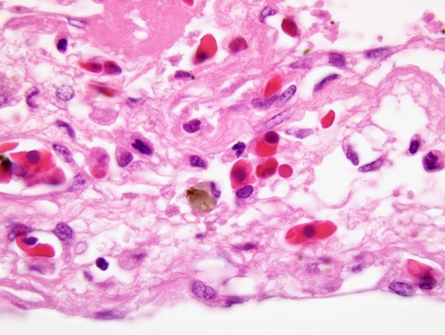
Microscopic examination of the lung consists of a hemorrhagic, necrotizing
bronchopneumonia with massive aerial mycelial growth within the airways. These affected airways are filled with
numerous degenerated heterophils admixed with macrophages, necrotic debris, and bacillary colonies. The fungal
organisms contain 3-5 μm chains of conidia, 4-7 μm dichotomous-branching, septate fungal hyphae. Airways are
markedly dilated and the mucosal epithelium is extensively ulcerative with hemorrhage, many heterophils and
macrophages are invading into the adjacent lung parenchyma. In these pneumonic areas, inflammation is associated
with the appearance of numerous birefringent crystals with radiating spokes; the morphology of these crystals is
suggestive of calcium oxalate. Histochemistry reveals many PAS-positive fungal hyphae scattered throughout the
necrotic foci of lungs, liver, and spleen, primarily along the margins of the necrotic lesion.
Morphologic Diagnosis:
Lung: Bronchopneumonia, necrogranulomatous and hemorrhagic, severe,
subacute to chronic, multiple, with intralesional fungal hyphae, oxalate crystals, necrotic vasculitis, and pleuritis,
crocodile (
Crocodylus niloticus), reptile.
Lab Results:
Escherichia coli and
Aeromonas sp. were isolated from the lungs and liver.
Condition:
Aspergillus sp.
Contributor Comment:
Clinically, oxalate is produced by a variety of fungi, including saprophytic (e.g.
Aspergillus niger and
A. flavus) and phytopathogenic species, and other sources including methoxyflurane, ethylene
glycol, large doses of ascorbic acid (vitamin C), primary hyperoxaluria (humans, cats, and tibetan spaniels),
pyridoxine (vitamin B6) deficiency, and plants (e.g.Â
Halogeton glomeratus, Sarcobatus vermiculatus, Rheum
rhaponticum, Oxalis cernua, Rumex sp., Russian vine, etc.). Several pathways have been described for oxalic acid
production, but the mechanism of oxalate crystal production is not known. In
Aspergillus niger, the organism
apparently possesses oxaloacetate acetylhydrolase (OAH), one of the enzymes of the tricarboxylic acid cycle, and
degrades oxaloacetate to oxalate by this route (i.e. oxaloacetate + H2O â oxalate + acetate). In humans, similar
morphological findings have been reported, with often localized deposition of calcium oxalate crystals around a
pulmonary
A. niger fungal ball.(1-3,5,6)
Aspergillus, a ubiquitous environmental organism, is an opportunistic pathogen in mammals and birds.Â
Aspergillus
fumigatus is the most commonly reported cause of aspergillosis in animals, whereas
A. flavus, A. terreus, A.
nidulans, and
A. niger are incriminated less frequently. In animals, localized deposition of massive numbers of
calcium oxalate crystals in tissues has been reported in association with
Aspergillus infections.(5,7-9) This is the
first report of pulmonary oxalosis in a crocodile.
JPC Diagnosis:
Lung: Pneumonia, necrohemorrhagic, multifocal to coalescing, severe, with pleuritis, myriad
fungal hyphae, and abundant anisotropic crystalline material (oxalate crystals).
Conference Comment:
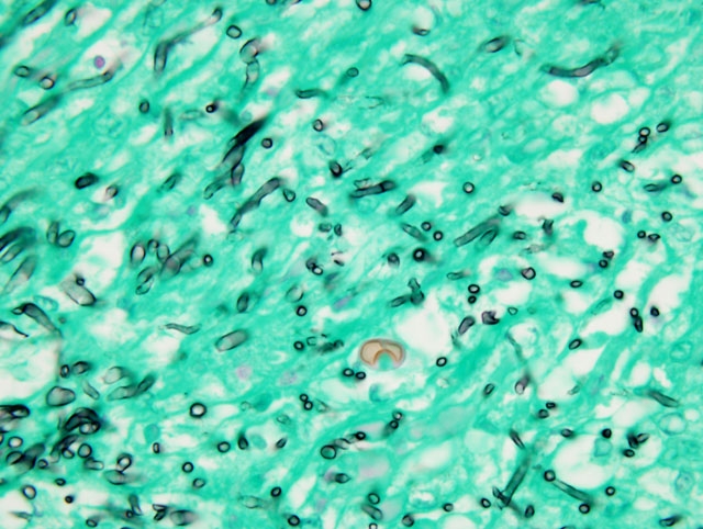
The GMS method highlights the fungal conidia and is helpful in demonstrating the other
hyphal characteristics described by the contributor; while these features, in combination with oxalate crystal
formation, are certainly suggestive of aspergillosis, the results of confirmatory testing (e.g. fungal culture) would be
particularly valuable for solidifying the specific etiologic diagnosis. Furthermore, while the morphology of the
prismatic crystalline material is consistent with oxalate crystals, histochemical stains (e.g. Yasues sliver nitraterubeanic
acid), though not performed, may be employed to definitively identify oxalate crystals.(5)
In domestic species, oxalate crystals are more commonly encountered in the renal tubules due to ethylene glycol
toxicosis in cats and dogs, or intoxication with one of the oxalate-containing plants listed above in sheep and cattle.
Interestingly, the rumen microflora have some capacity to degrade oxalate to bicarbonate and carbonates, conferring
sheep and cattle with a degree, albeit limited, of resistance to oxalosis; yet, remarkably, horses are far more resistant
to oxalate-induced nephrosis. Because oxalate chelates calcium, hypocalcemia is a characteristic finding in both
ethylene glycol toxicosis and oxalate toxicosis.(4)
References:
1. Blackmon JA:
Aspergillus niger. Am J Clin Pathol
76:506, 1981
2. Kauffman CA, Wilson KH, Schwartz DB: Necrotizing pulmonary aspergillosis with oxalosis. Mykosen
27:535-538, 1984
3. Kurrein F, Green GH, Rowles SL: Localized deposition of calcium oxalate around a pulmonary
Aspergillus
niger fungus ball. Am J Clin Pathol
64:556-563, 1975
4. Maxie MG, Newman SJ: Urinary system.Â
In: Jubb, Kennedy, and Palmers Pathology of Domestic Animals, ed.
Maxie MG, 5th ed., vol. 2, pp. 470-472. Elsevier Saunders, Philadelphia, PA, 2007
5. Muntz FH: Oxalate-producing pulmonary aspergillosis in an alpaca. Vet Pathol
36:631-632, 1999
6. Severo LC, Londero AT, Geyer GR, Picon PD: Oxalosis associated with an
Aspergillus niger fungus ball: report
of a case. Mycopathologia
73:29-31, 1981
7. Tham VL, Purcell DA: Fungal nephritis in a grey-headed albatross. J Wildl Dis
10:306-309, 1974
8. Wobeser G, Saunders JR: Pulmonary oxalosis in association with
Aspergillus niger infection in a great horned
owl (
Bubo virginianus). Avian Dis
19:388-392, 1975
9. Wyand DS, Langheinrich K, Helmboldt CF: Aspergillosis and renal oxalosis in a white-tailed deer. J Wildl Dis
7:52-56, 1971