Signalment:
3.5 year-old cowAnimal with a suppurative osteomyelitis of the right mandible
Gross Description:
In the right mandible, there was a hard mass 12 cm in diameter with ulceration of the adjacent gum. The mass was composed of many confluent fibrous nodules and several suppurative tracts. In the fibrous nodules, there were several cavities 1 mm to 1 cm in diameter containing variable amounts of a yellowish pus with many sulfur granules.
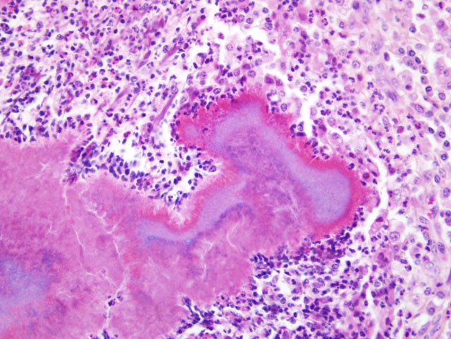
Histopathologic Description:
Most of the mandibular bone is replaced by a granulation tissue infiltrated by macrophages and plasma cells. The granulation tissue is surrounding many small abscesses with granules composed of large bacterial colonies (Gram-positive rod or coccoid-shaped and branching filamentous organisms) surrounded by radiating eosinophilic clubs (Splendore-Hoeppli material)
(Fig. 1-1). There is a zone of neutrophils around the granules, surrounded by many large macrophages and plasma cells.
Morphologic Diagnosis:
Chronic pyogranulomatous mandibular osteomyelitis, with large colonies of Gram-positive filamentous organisms
Condition:
Actinomyces bovis
Contributor Comment:
This pyogranulomatous mandibular osteomyelitis with the presence of colonies of Gram-positive branching filamentous organisms forming sulfur granules, is a good example of mandibular actinomycosis in cattle caused by
Actinomyces Bovis.
1, 2 The osteomyelitis would result from an extension of the infection of the gums or peridontium by the bacteria, following injury by foreign bodies or as a complication of periodontitis.
1 The excessive periosteal proliferation and the granulation tissue induced by the chronic inflammatory process, can cause a marked enlargement of the affected mandible (lumpy jaw).
JPC Diagnosis:
Bone; skeletal muscle; fibrous connective
tissue, right mandible (per contributor): Pyogranulomas,
multifocal to coalescing, with Splendore-Hoeppli
material and colonies of Gram-positive filamentous bacteria,
cow (
Bos taurus), bovine.
Conference Comment:
Actinomycetes are Grampositive,
non-acid-fast, branching filamentous rods.
They are facultative anaerobes and normal inhabitants of
the oral mucous membranes, tooth surfaces, and gastrointestinal
tract.
1,2 Actinomycosis, or lumpy jaw, is primarily
a disease of cattle, although it has been reported inhorses, pigs, deer, sheep and dogs.
2 Infections usually
are restricted to the bone of the mandible resulting in a
chronic suppurative and fibrosing osteomyelitis, although
infections have been reported to involve the maxilla, regional
lymph nodes or tongue.
2,3
Infection usually occurs secondary to trauma with subsequent
extension into the periosteum.
2 The normal architecture
of the mandible is progressively destroyed inciting
an extensive proliferative periosteal reaction.
2 The
purulent exudate may contain necrotic trabecular bone
(bone sand), or soft yellow granules containing mats of
tangled, filamentous bacteria and Splendore-Hoeppli material
(sulfur granules).
2
Residents at AFIP utilize the mnemonic YACS to develop
a differential diagnosis when large colonies of bacteria are present in hematoxylin and eosin stained sections.
YACS stands for:
Y
Yersinia sp.
A
Actinomyces sp., Actinobacillus sp. Arcanobacter sp.
C
Corynebacterium sp.
S
Staphylococcus sp., Streptococcus sp.
References:
1. Jones TC, Hunt RD, King NW: Diseases caused by
bacteria. In: Veterinary Pathology, eds. Jones TC, Hunt
RD, and King NW, 6th ed., pp. 482-484. Williams and
Wilkins, Baltimore, MA, 1997
2. Thompson K: Bones and joints. In: Jubb, Kennedy
and Palmers Pathology of Domestic Animals, ed. Maxie
MG, 5th ed., vol. 1, pp. 98-99. Elsevier Saunders, Philadelphia,
PA, 2007
3. Valentine BA, McGavin MD: Skeletal muscle. In:
Pathologic Basis of Veterinary Disease, eds. McGavin
MD, Zachary JF, 4th ed., p. 1020. Elsevier, St. Louis,
MO, 2007