Signalment:
Gross Description:
Histopathologic Description:
Morphologic Diagnosis:
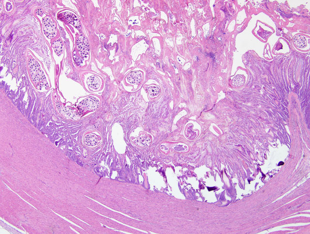
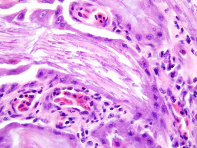
1. Ventriculus, ventriculitis, heterophilic and lymphocytic, multifocal, severe with koilin disruption and intralesional nematodes, bacteria and yeasts (some sections).
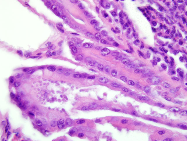
2. Proventriculus, proventriculitis, lymphocytic, multifocal, mild with intralesional yeasts (some sections) and apical protozoa (some sections).
Condition:
Contributor Comment:
Acuaria spp. have been reported in cases of finches with a history of diarrhea, lethargy and inappetence.(6) Spiruroids are active parasites of many finch species, especially the ground dwelling species of finch like long-tailed finches, and can be a significant source of mortality.(6) In addition to finches, A. skrjabini has been reported to infect and cause severe disease in sparrows from multiple regions of the world.(8) Fatal A. skrjabini infections with severe ventricular lesions have been reported in aviary finches in Australia and New Zealand.(8) In one population of aviary finches infected with A. skrajabini, there was a reported 66% mortality rate with severe gizzard lesions.(3) Other findings associated with Acuaria spp. include no gross lesions, embryonated eggs in intestinal smears, poor body condition, whole seeds in the intestinal tract, tapeworms in the intestine, and degeneration of the koilin with foci of bacteria.(3,4,8) In this population of finches, small nematodes rarely could be seen at necropsy beneath the koilin layer of the ventriculus.
Acuaria spp. have a two-host life cycle that requires an arthropod intermediate host, which may include beetles, sandhoppers and grasshoppers.(6) Treatment with oral levamisole or fenbendazole has been reported to reduce morbidity and mortality in affected birds,(6) providing supportive evidence that the disruption caused by these spiruroid gizzard worms is the primary cause of this disease syndrome.
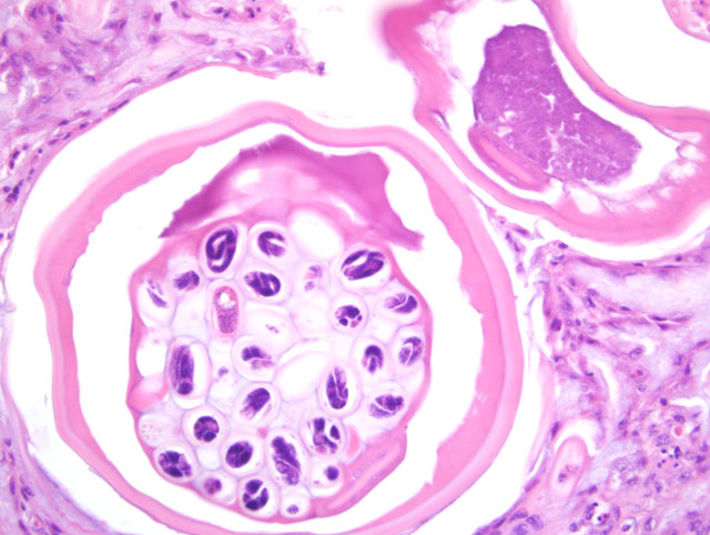
Speciation of Acuaria nematodes is difficult, although there are some features to distinguish A. skrjabini. Adult A. skrjabini parasites have seven pairs of postanal papillae in the male, and male cordons reach to and usually beyond the excretory pore.(5) The caudal alae of the male are wider anteriorly.(5) Females are characterized by thick shelled eggs approximately 40-43 x 23-24 um in the common trunk of uterus.(5) There are also distinguishing features between male and female worms; for example, female worms are about four times as long as the males, and transverse striations at mid body are at about 4 um intervals in the male while they are at 8 um intervals in the female.(5)
Yeast forms consistent with Macrorhabdus ornithogaster were also found in the proventriculus of five of the affected birds in this population. M. ornithogaster is a Gram positive, ascomycetous yeast which was originally classified as a bacterium. This organism has been reported in a variety of avian species, including finches, budgerigars and cockatiels. The yeasts are often found at the isthmus between the ventriculus and proventriculus, along the mucosal surface or within proventricular glands. Organisms isolated from chickens are infectious to young chicks, causing reduced weight gain and heterophilic inflammation of the isthmus of the proventriculus.(2)
Cryptosporidia are apicomplexan parasites that infect a wide range of hosts. Cryptosporidia occupy a unique intracellular but extracytoplasmic location on the apical surface of mucosal epithelial cells. Avian crytopsporidiosis can affect the epithelia of the respiratory tract, genitourinary tract, gastrointestinal tract or bursa of Fabricius. In birds, cryptosporidiosis has been reported in chickens, turkeys, quail, pheasants, peafowl, junglefowl, ducks, geese, parrots, finches, lovebirds, and budgerigars. Cryptosporidium meleagridis and C. baileyi are the species that infect turkeys and chickens, respectively. Two novel species, infecting black ducks and finches, have also been described. (7)
JPC Diagnosis:
1. Ventriculus: Ventriculitis, heterophilic and lymphocytic, multifocal, severe, with intramucosal spirurid eggs, larvae, and adults and numerous yeasts, etiology consistent with Acuaria species and Macrorhabdus ornithogaster, respectively.
2. Proventriculus: Proventriculitis, lymphocytic, multifocal, mild to moderate, with yeasts and many surface epithelial-associated protozoa, etiology consistent with Macrorhabdus ornithogaster and Cryptosporidium species, respectively.
Conference Comment:
During the conference, attendees first discussed the differential diagnosis for proventricular and ventricular nematodiasis, and like the contributor, narrowed the list to Acuaria sp. and Synhimatus (Dispharynx) sp. based on the histomorphology of the nematodes and favored Acuaria sp. because of the location. An example of the Synhimatus (Dispharynx) sp. in the proventriculus of a Northern bobwhite quail was reviewed in WSC 2008-2009, Conference 18, case II.
This case was reviewed in consultation with Dr. Christopher Gardiner, Consulting Parasitologist for the AFIPs Department of Veterinary Pathology, who emphasized that the presence of many small, thick-shelled, embryonated eggs is the most obvious indicator of a spirurid infection. However, the absence of characteristic spirurid eggs is not always sufficient to exclude a spirurid infection, because several spirurids (e.g. Draschia and Thalazia spp.) produce embryos lacking shells. Although there is substantial morphological variety among the spirurids, other features typically observed in members of the group include coelomyarian musculature, large intestines lined by uninucleate cuboidal to columnar cells that often have a brush border, large lateral chords that are often vacuolated, and eosinophilic fluid within the pseudocoelom.(1)
Particularly intriguing is the fact that both in this case and in reports in the literature(3,6,8) infection with Acuaria skrjabini manifests as an outbreak, despite its requirement for an intermediate host. In one aviary outbreak, a ceramic heat lamp attracted an infestation of cockroaches and earwigs, which were implicated as possible intermediate hosts. The authors noted that while finches of the Estrildidae family normally feed primarily on grass seeds, they transition to a greater dietary intake of insects during the breeding season, which may account for the nearly simultaneous onset of disease in a number of birds. Death of affected birds was attributed to starvation due to maldigestion caused by ventricular dysfunction.(3)
The contributor alluded to the original misclassification of M. ornithogaster as a bacterium (based on Gram staining characteristics), giving rise to the misnomer megabacterium, by which it was formerly known. M. ornithogaster survives only in a microaerophilic environment in a narrow pH range of 3-4, consistent with its peculiar environmental niche at the isthmus of the proventriculus and ventriculus of birds.(2) Although many birds appear asymptomatically infected, the yeast has been implicated in three specific avian disease syndromes: 1) chronic wasting disease in budgerigars, canaries, and finches; 2) acute hemorrhagic gastritis in budgerigars and parrotlets; and 3) reduced feed conversion in experimentally-infected chickens.(2) The clinical significance of the organism in the present case is unclear, but most conference participants interpreted it as an incidental finding, and certainly less important than the spirurids discussed above. By contrast, because they are so numerous and associated with areas of proventricular inflammation, the cryptosporidia in this case are considered clinically significant.
References:
2. Hannafusa Y, Bradley A, Tomaszewski EE, Libal MC, Phalen DN: Growth and metabolic characterization of Macrorhabdus ornithogaster. J Vet Diagn Invest 19:256-265, 2007
3. Hindmarsh M, Ward K: Mortality of finches (family Estrildidae) caused by Acuaria skrjabini. Aust Vet J 70:451-452, 1993
4. Mason PC, Hodgkinson NL, McAllum HJ: Acuaria skrjabini Ozerska, 1926 in a caged finch. N Z Vet J 26:131-132, 1978
5. Mawson PM: The genus Acuaria Bremser (Nematoda: Spirurida) in Australia. Trans R Soc S Aust 96:139-147, 1972
6. McOrist S, Barton NJ, Black DJ: Acuaris skrjabini infection of the gizzard of finches. Avian Dis 26:957-960, 1982
7. Morgan UM, Monis PT, Xiao L, Limor J, Sulaiman I, Raidal S, O'Donoghue P, Gasser R, Murray A, Fayer R, Blagburn BL, Lal AA, Thompson RC: Molecular and phylogenetic characterisation of Cryptosporidium from birds. Int J Parasitol 31:289-296, 2001
8. Sato H, Osanai A, Kamiya H, Une Y: Gizzard spirurid nematode Acuaria skrjabini in Japanese tree sparrows and a gray starling from Tokyo. J Vet Med Sci 67:607-609, 2005